Rudolph MG
Rudolph MG
Rudolph MG
You also want an ePaper? Increase the reach of your titles
YUMPU automatically turns print PDFs into web optimized ePapers that Google loves.
Annu. Rev. Immunol. 2006.24:419-466. Downloaded from arjournals.annualreviews.org<br />
by CAMBRIDGE UNIVERSITY on 04/04/10. For personal use only.<br />
structure of human CD4 is also known (110),<br />
superposition of the MHC-proximal CD4 domains<br />
1 and 2 permits assembly of a complete<br />
class II TCR/pMHC/CD4 model that suggests<br />
a V-shape with the T cell membraneproximal<br />
ends of the TCR and CD4<br />
separated by around 100 ˚A. This separation<br />
would exclude direct TCR/CD4 interactions,<br />
but leaves ample room for binding of<br />
CD3.<br />
CD3 SIGNALING MODULES<br />
CD3 consists of subunits δ, ε, γ, and ζ<br />
that noncovalently associate to form CD3ζζ<br />
homodimers and CD3εδ and CD3εγ heterodimers<br />
(111, 112). Whereas sustained T<br />
cell responses rely on coreceptor binding and<br />
TCR aggregation (113–116), early TCR signaling<br />
is independent of these events but may<br />
instead rely on conformational changes in the<br />
CD3ε subunit (84). In addition, stable cell<br />
surface expression and normal development<br />
of αβ TCRs rely on the presence of the<br />
CD3 components (117–119). Sequence comparisons<br />
predicted that the extracellular domains<br />
of the CD3ε- and CD3γ-chains would<br />
adopt an immunoglobulin fold (120). A cavity<br />
formed by the FG loop in the Cβ domain<br />
of αβ TCRs was suggested to host a binding<br />
site for such an Ig-domain (9), but other<br />
studies reported the dispensability of any Igdomain<br />
residues in CDεδ for TCR α-chain<br />
binding, limiting the key residues to the conserved<br />
charged transmembrane residues on<br />
CD3 and the TCR (121). The first insights<br />
into these signaling modules came from an<br />
NMR structure in which the extracellular domains<br />
of the mouse CD3ε and -γ subunits<br />
that lacked the conserved RxCxxCxE stalk region<br />
motif, considered important for dimerization,<br />
were converted to a single-chain<br />
format by a 26-residue linker that ensured<br />
close proximity during folding from inclusion<br />
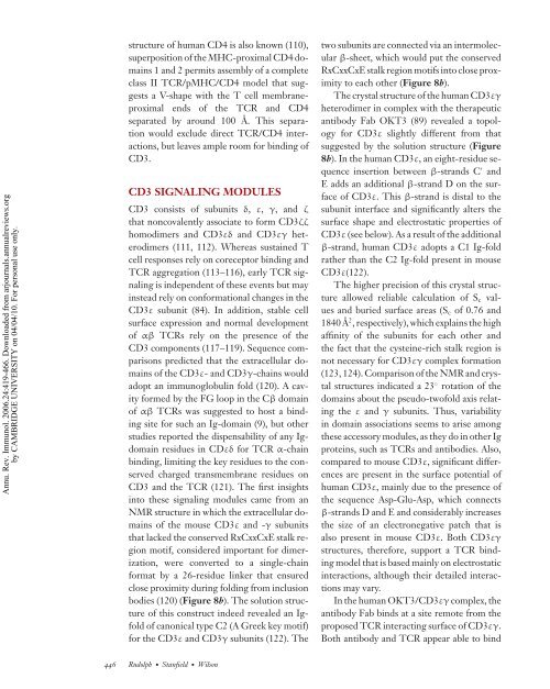
bodies (120) (Figure 8b). The solution structure<br />
of this construct indeed revealed an Igfold<br />
of canonical type C2 (A Greek key motif)<br />
for the CD3ε and CD3γ subunits (122). The<br />
446 <strong>Rudolph</strong>· Stanfield· Wilson<br />
two subunits are connected via an intermolecular<br />
β-sheet, which would put the conserved<br />
RxCxxCxE stalk region motifs into close proximity<br />
to each other (Figure 8b).<br />
The crystal structure of the human CD3εγ<br />
heterodimer in complex with the therapeutic<br />
antibody Fab OKT3 (89) revealed a topology<br />
for CD3ε slightly different from that<br />
suggested by the solution structure (Figure<br />
8b). In the human CD3ε, an eight-residue sequence<br />
insertion between β-strands C ′ and<br />
E adds an additional β-strand D on the surface<br />
of CD3ε. This β-strand is distal to the<br />
subunit interface and significantly alters the<br />
surface shape and electrostatic properties of<br />
CD3ε (see below). As a result of the additional<br />
β-strand, human CD3ε adopts a C1 Ig-fold<br />
rather than the C2 Ig-fold present in mouse<br />
CD3ε(122).<br />
The higher precision of this crystal structure<br />
allowed reliable calculation of Sc values<br />
and buried surface areas (Sc of 0.76 and<br />
1840 ˚A 2 , respectively), which explains the high<br />
affinity of the subunits for each other and<br />
the fact that the cysteine-rich stalk region is<br />
not necessary for CD3εγ complex formation<br />
(123, 124). Comparison of the NMR and crystal<br />
structures indicated a 23 ◦ rotation of the<br />
domains about the pseudo-twofold axis relating<br />
the ε and γ subunits. Thus, variability<br />
in domain associations seems to arise among<br />
these accessory modules, as they do in other Ig<br />
proteins, such as TCRs and antibodies. Also,<br />
compared to mouse CD3ε, significant differences<br />
are present in the surface potential of<br />
human CD3ε, mainly due to the presence of<br />
the sequence Asp-Glu-Asp, which connects<br />
β-strands D and E and considerably increases<br />
the size of an electronegative patch that is<br />
also present in mouse CD3ε. Both CD3εγ<br />
structures, therefore, support a TCR binding<br />
model that is based mainly on electrostatic<br />
interactions, although their detailed interactions<br />
may vary.<br />
In the human OKT3/CD3εγ complex, the<br />
antibody Fab binds at a site remote from the<br />
proposed TCR interacting surface of CD3εγ.<br />
Both antibody and TCR appear able to bind


