‹nmeli Hastalarda Yaflflam Kalitesini Etkileyen ... - FTR Dergisi
‹nmeli Hastalarda Yaflflam Kalitesini Etkileyen ... - FTR Dergisi
‹nmeli Hastalarda Yaflflam Kalitesini Etkileyen ... - FTR Dergisi
Create successful ePaper yourself
Turn your PDF publications into a flip-book with our unique Google optimized e-Paper software.
181<br />
23. Ulusal Fiziksel T›p ve Rehabilitasyon Kongresi / 23 rd National Physical Medicine & Rehabilitation Congress<br />
Türk Fiz Rehab Derg 2011:57Özel Say›; 1-334 /Turk J Phys Med Rehab 2011:57Suppl; 1-334<br />
P-054<br />
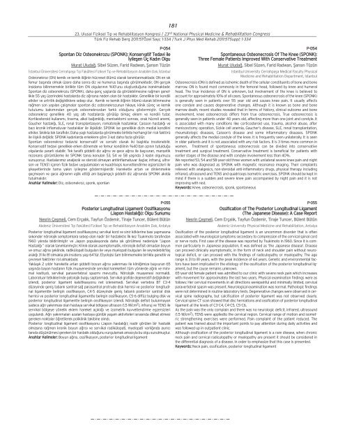
Spontan Diz Osteonekrozu (SPONK): Konservatif Tedavi ile<br />
‹yileflen Üç Kad›n Olgu<br />
Murat Uluda¤, Sibel Süzen, Farid Radwan, fians›n Tüzün<br />
‹stanbul Üniversitesi Cerrahpafla T›p Fakültesi Fiziksel T›p ve Rehabilitasyon Anabilim Dal›, ‹stanbul<br />
Osteonekroz (ON) kemik ve kemik ili¤inin hücresel ölümü olarak tan›mlanmaktad›r. ON en s›k<br />
femur bafl›nda olmak üzere daha sonra diz ve humerus bafl›nda görülmektedir. ON gerçek<br />
insidans› bilinmemekle birlikte tüm ON olgular›n›n %10’unu oluflturdu¤una inan›lmaktad›r.<br />
Spontan diz osteonekrozu (SPONK), daha genç yafllarda da görülebilmesine ra¤men genellikle<br />
55 yafl üzerindeki hastalarda diz a¤r›s›na neden olan bir hastal›kt›r. Genellikle bir kondili<br />
etkiler ve artritik de¤iflikliklere sebep olur. Kemik ve kemik ili¤inin ölümü olarak bilinmesine<br />
ra¤men son yap›lan çal›flmalar spontan diz osteonekrozunun hikaye, klinik süreç ve kemik<br />
tutulumu bak›m›ndan gerçek osteonekrozdan farkl› oldu¤unu göstermifltir. Gerçek<br />
osteonekroz genellikle 40 yafl alt› hastalarda görülüp birkaç eklem ve kondili tutar.<br />
Kortikosteroid kullan›m›, travma, alkol ba¤›ml›l›¤›, menisektomi sonras›, orak hücreli anemi,<br />
Gaucher hastal›¤›, SLE, renal transplantasyon, romatolojik hastal›klar, Caisson hastal›¤› ve<br />
baz› kronik inflamatuvar hastal›klar ile iliflkilidir. SPONK ise genellikle dizin medial kondilini<br />
etkiler. S›kl›kla tek tarafl›d›r. Daha yafll› hastalarda görülmekle birlikte herhangi bir risk faktörü<br />
ile iliflkili de¤ildir. SPONK kad›nlarda erkeklere göre 3 kat daha fazla görülür.<br />
Spontan osteonekroz tedavisi konservatif ve cerrahi olarak iki bafll›kta incelenebilir.<br />
Konservatif tedavi genellikle erken dönemde ve femur kondilinin %40’dan az›n›n tutuldu¤u<br />
olgularda yararl› olabilir. Tek tarafl› fliddetli diz a¤r›s› ve gece a¤r›s› ile baflvuran, manyetik<br />
rezonans görüntüleme ile SPONK tan›s› konulan 53, 54 ve 58 yafl›nda 3 kad›n olgumuzu<br />
sunuyoruz. Hastalar›m›z analjezik ve steroid olmayan antiinflamatuvar ilaçlar, infraruj, ultrason<br />
ve TENS’i içeren fizik tedavi uygulamalar› ve kuadriseps kuvvetlendirme egzersizleri ile<br />
flikayetlerinde tama yak›n iyileflme göstermifllerdir. Hareketle artan ve dinlenmekle<br />
geçmeyen ve gece a¤r›s›n›n efllik etti¤i ani bafllang›çl› fliddetli diz a¤r›s›nda SPONK ak›lda<br />
tutulmal›d›r.<br />
Anahtar Kelimeler: Diz, osteonekroz, sponk, spontan<br />
P-055<br />
Posterior Longitudinal Ligament Ossifikasyonu<br />
(Japon Hastal›¤›): Olgu Sunumu<br />
Nesrin Çeflmeli, Cem Erçal›k, Tayfun Özdemir, Tiraje Tuncer, Bülent Bütün<br />
Akdeniz Üniversitesi T›p Fakültesi Fiziksel T›p ve Rehabilitasyon Anabilim Dal›, Antalya<br />
Posterior longitudinal ligament ossifikasyonu; servikal kord ve sinir köklerine bas› yapmas›na<br />
sekonder nörolojik semptomlara sebep olan nadir bir hastal›kt›r. ‹lk kez Tsukimoto taraf›ndan<br />
1960 y›l›nda bildirilmifltir ve Japon populasyonda daha s›k görülmesi nedeniyle ‘‘Japon<br />
Hastal›¤› ’’ olarak tan›mlanm›flt›r. Klinik olarak asemptomatik, nörolojik defisit olmadan boyun<br />
ve omuz a¤r›s› fleklinde, radikülopati bulgular› ile veya myelopati bulgular› ile seyredebilir.Yafl<br />
aral›¤› 31 ile 81 olmakla pik insidens yafl› 64’tür. Etyolojisi tam bilinmemekle birlikte genetik ve<br />
çevresel faktörler rol almaktad›r.<br />
Yaklafl›k 2 y›ld›r hareketle artan fliddetli boyun a¤r›s› yak›nmas› ile klini¤imize baflvuran 65<br />
yafl›nda bayan hastan›n fizik muayenesinde servikal hareketleri tüm yönlerde a¤r›l› ve minimal<br />
k›s›tl›yd›, servikal paravertebral spazm› mevcuttu. Nörolojik muayenesi normaldi.<br />
Laboratuar tetkiklerinde patolojik bulgu saptanmad›. Servikal grafide dejeneratif de¤ifliklikler<br />
izlendi, posterior ligament kalsifikasyonu net izlenemedi. Servikal vertebra BT C3-4<br />
düzeyinde genifl tabanl› santral-sa¤ parasantral protrude disk hernisi ve posterior longitudinal<br />
ligamentte belirgin ossifikasyon, C4-5 düzeyinde genifl tabanl› posterior santral disk<br />
hernisi ve posterior longitudinal ligamentte belirgin ossifikasyon, C5-6 diffüz bulging disk ve<br />
posterior longitudinal ligamentte belirgin ossifikasyon izlendi. Nörolojik defisit bulunmayan<br />
sadece a¤r› yak›nmas› olan hastaya servikal bölgeye ultrason (1,5 W/cm2 ), infraruj ve TENS ile<br />
servikal bölgeye yönelik eklem hareket aç›kl›¤› ve izometrik kuvvetlendirme egzersizleri<br />
uyguland›. A¤r› yak›nmalar› azalan hastaya günlük yaflam aktiviteleri s›ras›nda dikkat etmesi<br />
gereken noktalar ö¤retilerek poliklinik takibine al›nd›.<br />
Posterior longitudinal ligament ossifikasyonu (Japon hastal›¤›); nadir görülen bir hastal›k<br />
olmas›na ra¤men kronik boyun a¤r›s› ve servikal radikülopati, myelopati varl›¤›nda ay›r›c›<br />
tan›da düflünülmesi gereken bir hastal›k oldu¤unu vurgulamak amac›yla bu olgu sunulmufltur.<br />
Anahtar Kelimeler: Boyun a¤r›s›, ossifikasyon, posterior longitudinal ligament<br />
P-054<br />
Spontaneous Osteonecrosis Of The Knee (SPONK):<br />
Three Female Patients Improved With Conservative Treatment<br />
Murat Uluda¤, Sibel Süzen, Farid Radwan, fians›n Tüzün<br />
Istanbul University Cerrahpafla Medical Faculty Physical<br />
Medicine and Rehabilitation Department, Istanbul<br />
Osteonecrosis (ON) is defined as ischemic death of the cellular constituents of bone and bone<br />
marrow. ON is found most commonly in the femoral head, followed by knee and humeral<br />
head. The true incidence of ON is unknown, but involvement of the knee is believed to<br />
account for approximately 10% of all cases. Spontaneous osteonecrosis of the knee (SPONK)<br />
is generally seen in patients over 55 year old and causes knee pain. It usually affects<br />
one condyle and causes degenerative changes. Although it is known as bone and bone<br />
marrow death, recent studies revealed that in terms of history, clinical outcome and bone<br />
involvement, knee osteonecrosis differs from true osteonecrosis. True osteonecrosis is<br />
generally seen in patients under 40 years old, affecting more than one joint and condyle. It<br />
is associated with many conditions like corticosteroid use, trauma, alcohol abuse, after<br />
meniscectomy operation, Sickle cell anemia, Gaucher’s disease, SLE, renal transplantation,<br />
rheumatologic diseases, Caisson’s disease and some inflammatory diseases. SPONK<br />
generally affects the medial condyle of the knee. It is frequently seen unilaterally. It is seen<br />
in older patients and it is not associated with any risk factors. It is 3 times more common in<br />
women. Treatment of spontaneous osteonecrosis can be divided into conservative<br />
treatment and surgical treatment. Conservative treatment is beneficial for patients with<br />
earlier stages of the disease and with condyle involvement less than 40%.<br />
We reported 53, 54 and 58-year-old three women with unilateral severe knee pain and night<br />
pain who was diagnosed as SPONK with magnetic resonance imaging. Their complaints<br />
relieved with analgesics, non-steroidal anti-inflammatory drugs, physical therapy including<br />
infrared, ultrasound and TENS and quadriceps isometric exercises. SPONK should be kept in<br />
mind if there is a sudden and severe knee pain accompanied by night pain and it is not<br />
improving with rest.<br />
Keywords: Knee, osteonecrosis, sponk, spontaneous<br />
P-055<br />
Ossification of The Posterior Longitudinal Ligament<br />
(The Japanese Disease): A Case Report<br />
Nesrin Çeflmeli, Cem Erçal›k, Tayfun Özdemir, Tiraje Tuncer, Bülent Bütün<br />
Akdeniz University Physical Medicine and Rehabilitation, Antalya<br />
Ossification of the posterior longitudinal ligament is an uncommon disorder that is often<br />
associated with neurological symptoms secondary to compression of the cervical spinal cord<br />
or nerve roots. First case of the disease was reported by Tsukimoto in 1960. Since it is common<br />
particularly in Japanese population, It was defined as 'the Japanese disease'. Disease<br />
can proceed clinically asymptomatic, in the form of neck and shoulder pain without neurological<br />
deficit, or can proceed with the findings of radiculopathy or myelopathy. The age<br />
range is 31 to 81 years, with the peak incidence of 64 years. Genetic and environmental factors<br />
have been implicated in the etiology of the ossification of the posterior longitudinal ligament,<br />
but the cause remains unknown.<br />
65-year-old female patient was admitted to our clinic with severe neck pain which increases<br />
with movement for approximately the last two years. Physical examination findings were as<br />
follows: Her cervical movements in all directions werepainful and minimally limited, cervical<br />
paravertebral spasm was present. Neurological examination was normal. Pathologic findings<br />
were not determined in routine laboratory tests. Degenerative changes were observed in cervical<br />
spine radiography, but calcification of posterior ligament was not observed clearly.<br />
Cervical spine CT scan showed that disc herniations and ossification of posterior longitudinal<br />
ligament at the levels of C3-C4, C4-C5, C5-C6.<br />
As the pain was the only complain and there was no neurologic deficit, infrared, ultrasound<br />
(1.5 W/cm 2 ), TENS were appliedto the cervical region. Cervical range of motion and isometric<br />
strengthening exercises were performed. Pain complaint of the patient reduced. The<br />
patient was trained about the important points to pay attention during daily activities and<br />
was followed up in outpatient clinic.<br />
Although ossification of the posterior longitudinal ligament is a rare disease, when chronic<br />
neck pain and cervical radiculopathy or myelopathy are present it should be considered in<br />
the differential diagnosis of a disease. In order to emphasize that this case is presented.<br />
Keywords: Neck pain, ossification, posterior longitudinal ligament


