Zemes un vides zinātnes Earth and Environment Sciences - Latvijas ...
Zemes un vides zinātnes Earth and Environment Sciences - Latvijas ...
Zemes un vides zinātnes Earth and Environment Sciences - Latvijas ...

Create successful ePaper yourself
Turn your PDF publications into a flip-book with our unique Google optimized e-Paper software.
140<br />
ADVANCES IN PALAEOICHTHYOLOGY<br />
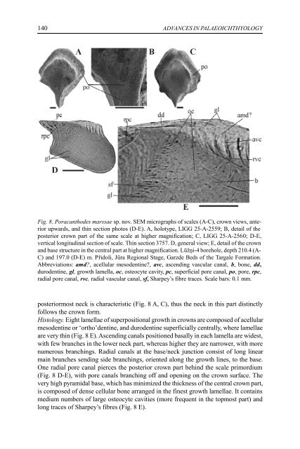
Fig. 8. Poracanthodes marssae sp. nov. SEM micrographs of scales (A-C), crown views, anterior<br />
upwards, <strong>and</strong> thin section photos (D-E). A, holotype, LIGG 25-A-2559; B, detail of the<br />
posterior crown part of the same scale at higher magnification; C, LIGG 25-A-2560; D-E,<br />
vertical longitudinal section of scale. Thin section 3757. D, general view; E, detail of the crown<br />
<strong>and</strong> base structure in the central part at higher magnification. Lūžņi-4 borehole, depth 210.4 (A-<br />
C) <strong>and</strong> 197.0 (D-E) m. Přidoli, Jūra Regional Stage, Garzde Beds of the Targale Formation.<br />
Abbreviations: amd?, acellular mesodentine?, avc, ascending vascular canal, b, bone, dd,<br />
durodentine, gl, growth lamella, oc, osteocyte cavity, pc, superficial pore canal, po, pore, rpc,<br />
radial pore canal, rvc, radial vascular canal, sf, Sharpey’s fibre traces. Scale bars: 0.1 mm.<br />
posteriormost neck is characteristic (Fig. 8 A, C), thus the neck in this part distinctly<br />
follows the crown form.<br />
Histology. Eight lamellae of superpositional growth in crowns are composed of acellular<br />
mesodentine or ‘ortho’dentine, <strong>and</strong> durodentine superficially centrally, where lamellae<br />
are very thin (Fig. 8 E). Ascending canals positioned basally in each lamella are widest,<br />
with few branches in the lower neck part, whereas higher they are narrower, with more<br />
numerous branchings. Radial canals at the base/neck j<strong>un</strong>ction consist of long linear<br />
main branches sending side branchings, oriented along the growth lines, to the base.<br />
One radial pore canal pierces the posterior crown part behind the scale primordium<br />
(Fig. 8 D-E), with pore canals branching off <strong>and</strong> opening on the crown surface. The<br />
very high pyramidal base, which has minimized the thickness of the central crown part,<br />
is composed of dense cellular bone arranged in the finest growth lamellae. It contains<br />
medium numbers of large osteocyte cavities (more frequent in the topmost part) <strong>and</strong><br />
long traces of Sharpey’s fibres (Fig. 8 E).