BIOLOGY IN FOCUS
BIOLOGY IN FOCUS
BIOLOGY IN FOCUS

Create successful ePaper yourself
Turn your PDF publications into a flip-book with our unique Google optimized e-Paper software.
CELLS AND THE CELL THEORY<br />
images are produced using a beam<br />
of electrons—electrons that are<br />
made to behave like light (waves).<br />
In 1928, Ernst Ruska and his<br />
supervisor Max Kroll built the first<br />
electron microscope, but it only<br />
had a magnification of 17×. Ruska<br />
continued working on the device<br />
and by 1933 he had built the first<br />
transmission electron microscope<br />
that could magnify up to 12 000×.<br />
Ruska’s team continued working<br />
on the electron microscope during<br />
the second world war, achieving a<br />
magnification of one million times.<br />
■ The basic principle of the<br />
transmission electron microscope<br />
is similar to that of the compound<br />
light microscope, except that<br />
the energy source transmitted<br />
through the specimen is a beam of<br />
electrons instead of a beam of light.<br />
Modifications to the design have had<br />
to be made because electrons do not<br />
normally travel in a manner similar<br />
to light, but bounce off anything that<br />
they hit, such as air. The electrons<br />
must therefore pass through the<br />
specimen in a vacuum, making it<br />
possible to view only non-living,<br />
preserved tissue. The electrons are<br />
focused by electron magnets, rather<br />
than by glass lenses, and the image is<br />
produced on a screen where it shows<br />
up as fluorescence, or it may be<br />
projected onto a photographic plate.<br />
■ The invention of the scanning<br />
electron microscope followed in<br />
1955. The electron beam causes the<br />
specimen to emit its own electrons,<br />
producing a three-dimensional<br />
image (but it has a low resolution).<br />
The picture on the front cover of this<br />
textbook was produced by a modern<br />
day, scanning electron microscope.<br />
Advantages<br />
The main advantage of the transmission<br />
electron microscope is the high<br />
magnification and resolution which<br />
show an enormous amount of detail.<br />
The electron microscope reveals<br />
structures at not only the cellular level,<br />
but also the sub-cellular level. Many<br />
parts of cells (organelles) were seen<br />
for the first time after the invention<br />
of the electron microscope. Other<br />
parts previously seen with the light<br />
microscope can be seen in far greater<br />
detail, providing increased knowledge<br />
of their internal structure. This has led<br />
to an understanding of their functions<br />
in cells.<br />
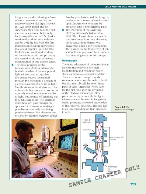
source of<br />
electrons<br />
Figure 1.8 The<br />
electron microscope<br />
specimen<br />
eye<br />
electromagnetic<br />
lenses<br />
final image<br />
on<br />
photographic<br />
plate or<br />
screen<br />
SAMPLE CHAPTER ONLY<br />
75