BIOLOGY IN FOCUS
BIOLOGY IN FOCUS
BIOLOGY IN FOCUS

You also want an ePaper? Increase the reach of your titles
YUMPU automatically turns print PDFs into web optimized ePapers that Google loves.
PATTERNS <strong>IN</strong> NATURE<br />
TR<br />
General resources<br />
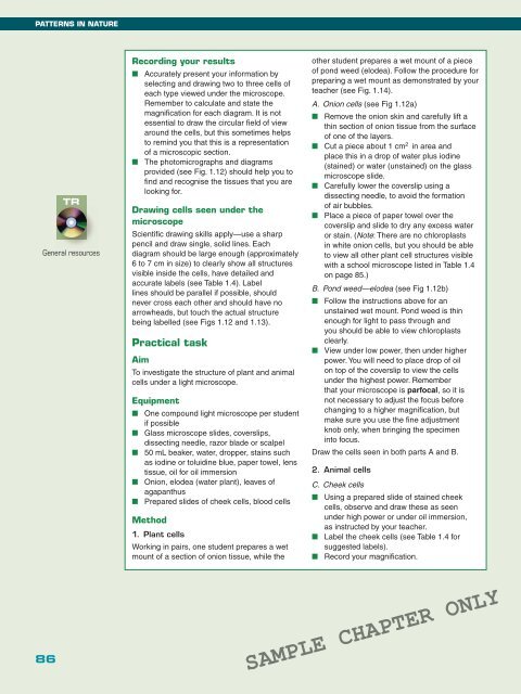
Recording your results<br />
■ Accurately present your information by<br />
selecting and drawing two to three cells of<br />
each type viewed under the microscope.<br />
Remember to calculate and state the<br />
magnifi cation for each diagram. It is not<br />
essential to draw the circular fi eld of view<br />
around the cells, but this sometimes helps<br />
to remind you that this is a representation<br />
of a microscopic section.<br />
■ The photomicrographs and diagrams<br />
provided (see Fig. 1.12) should help you to<br />
fi nd and recognise the tissues that you are<br />
looking for.<br />
Drawing cells seen under the<br />
microscope<br />
Scientifi c drawing skills apply—use a sharp<br />
pencil and draw single, solid lines. Each<br />
diagram should be large enough (approximately<br />
6 to 7 cm in size) to clearly show all structures<br />
visible inside the cells, have detailed and<br />
accurate labels (see Table 1.4). Label<br />
lines should be parallel if possible, should<br />
never cross each other and should have no<br />
arrowheads, but touch the actual structure<br />
being labelled (see Figs 1.12 and 1.13).<br />
Practical task<br />
Aim<br />
To investigate the structure of plant and animal<br />
cells under a light microscope.<br />
Equipment<br />
■ One compound light microscope per student<br />
if possible<br />
■ Glass microscope slides, coverslips,<br />
dissecting needle, razor blade or scalpel<br />
■ 50 mL beaker, water, dropper, stains such<br />
as iodine or toluidine blue, paper towel, lens<br />
tissue, oil for oil immersion<br />
■ Onion, elodea (water plant), leaves of<br />
agapanthus<br />
■ Prepared slides of cheek cells, blood cells<br />
Method<br />
1. Plant cells<br />
Working in pairs, one student prepares a wet<br />
mount of a section of onion tissue, while the<br />
other student prepares a wet mount of a piece<br />
of pond weed (elodea). Follow the procedure for<br />
preparing a wet mount as demonstrated by your<br />
teacher (see Fig. 1.14).<br />
A. Onion cells (see Fig 1.12a)<br />
■ Remove the onion skin and carefully lift a<br />
thin section of onion tissue from the surface<br />
of one of the layers.<br />
■ Cut a piece about 1 cm 2 in area and<br />
place this in a drop of water plus iodine<br />
(stained) or water (unstained) on the glass<br />
microscope slide.<br />
■ Carefully lower the coverslip using a<br />
dissecting needle, to avoid the formation<br />
of air bubbles.<br />
■ Place a piece of paper towel over the<br />
coverslip and slide to dry any excess water<br />
or stain. (Note: There are no chloroplasts<br />
in white onion cells, but you should be able<br />
to view all other plant cell structures visible<br />
with a school microscope listed in Table 1.4<br />
on page 85.)<br />
B. Pond weed—elodea (see Fig 1.12b)<br />
■ Follow the instructions above for an<br />
unstained wet mount. Pond weed is thin<br />
enough for light to pass through and<br />
you should be able to view chloroplasts<br />
clearly.<br />
■ View under low power, then under higher<br />
power. You will need to place drop of oil<br />
on top of the coverslip to view the cells<br />
under the highest power. Remember<br />
that your microscope is parfocal, so it is<br />
not necessary to adjust the focus before<br />
changing to a higher magnifi cation, but<br />
make sure you use the fi ne adjustment<br />
knob only, when bringing the specimen<br />
into focus.<br />
Draw the cells seen in both parts A and B.<br />
2. Animal cells<br />
C. Cheek cells<br />
■ Using a prepared slide of stained cheek<br />
cells, observe and draw these as seen<br />
under high power or under oil immersion,<br />
as instructed by your teacher.<br />
■ Label the cheek cells (see Table 1.4 for<br />
suggested labels).<br />
■ Record your magnifi cation.<br />
86<br />
SAMPLE CHAPTER ONLY