BIOLOGY IN FOCUS
BIOLOGY IN FOCUS
BIOLOGY IN FOCUS

Create successful ePaper yourself
Turn your PDF publications into a flip-book with our unique Google optimized e-Paper software.
CELLS AND THE CELL THEORY<br />
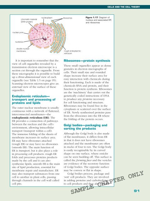
chromatin material<br />
(DNA protein)<br />
nucleoplasm<br />
ribosomes<br />
Figure 1.17 Diagram of<br />
nucleus and associated ER<br />
and ribosomes<br />
nuclear pore<br />
nucleolus<br />
double nuclear<br />
membrane<br />
rough endoplasmic<br />
reticulum<br />
It is important to remember that the<br />
view of cell organelles revealed by a<br />
transmission electron microscope is a<br />
section cut through the organelle. From<br />
these micrographs it is possible to build<br />
up a three-dimensional view of each<br />
organelle (see Table 1.5 on page 95).<br />
Scanning electron microscopes give an<br />
external view of the surface of these<br />
organelles.<br />
Endoplasmic reticulum—<br />
transport and processing of<br />
proteins and lipids<br />
The outer nuclear membrane is usually<br />
continuous with a network of flattened,<br />
interconnected membranes—the<br />
endoplasmic reticulum (ER). The<br />
ER provides a connection of pathways<br />
between the nucleus and the cell’s<br />
environment, allowing intracellular<br />
transport (transport within a cell).<br />
The immense folding of the sheets of<br />
membrane increases its surface area.<br />
ER may have ribosomes attached<br />
(rough ER) or may have no ribosomes<br />
(smooth ER). The main function of<br />
ER is transport, but it also plays a role<br />
in processing cell products: rough ER<br />
folds and processes proteins products<br />
made by the cell and it can also<br />
synthesise lipids; smooth ER is the main<br />
site of lipid production, essential for<br />
membrane repair and manufacture. ER<br />
may also transport substances from one<br />
cell to another in plant cells, passing<br />
through channels in the cell wall called<br />
cell pits.<br />
Ribosomes—protein synthesis<br />
These small organelles appear as dense<br />
granules in electron micrographs of<br />
cells. Their small size and rounded<br />
shape increase their surface area for<br />
easy interaction with chemicals during<br />
their functioning. Each is made of the<br />
chemicals RNA and protein, and their<br />
function is protein synthesis. Ribosomes<br />
are the ‘machinery’ that carries out the<br />
genetically coded instructions of DNA<br />
to produce any proteins necessary<br />
for cell functioning and structure.<br />
Ribosomes may be found free in the<br />
cytoplasm or scattered over the surface<br />
of ER. Newly synthesised proteins pass<br />
from the ribosomes into the ER where<br />
the folding of the protein occurs.<br />
Golgi bodies—packaging and<br />
sorting the products<br />
Although the Golgi body is also made<br />
of flat membranes, it differs from ER<br />
in that it does not have ribosomes<br />
attached and the membranes are often<br />
in stacks of four to ten. The Golgi body<br />
is easily recognisable by its curved<br />
shape on one surface, where vesicles<br />
can be seen budding off. This surface is<br />
called the forming face and the vesicles<br />
are evidence of the secretory function<br />
of Golgi bodies. The opposite surface<br />
may be convex or flat in shape.<br />
Golgi bodies process, package and<br />
‘sort’ cell products. They are involved<br />
in adding proteins and carbohydrates<br />
to cell products and they also provide<br />
SAMPLE CHAPTER ONLY<br />
91