BIOLOGY IN FOCUS
BIOLOGY IN FOCUS
BIOLOGY IN FOCUS

You also want an ePaper? Increase the reach of your titles
YUMPU automatically turns print PDFs into web optimized ePapers that Google loves.
CELLS AND THE CELL THEORY<br />
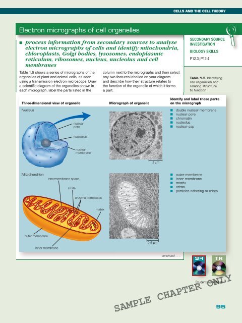
Electron micrographs of cell organelles<br />
■ process information from secondary sources to analyse<br />
electron micrographs of cells and identify mitochondria,<br />
chloroplasts, Golgi bodies, lysosomes, endoplasmic<br />
reticulum, ribosomes, nucleus, nucleolus and cell<br />
membranes<br />
Table 1.5 shows a series of micrographs of the<br />
organelles of plant and animal cells, as seen<br />
using a transmission electron microscope. Draw<br />
a scientifi c diagram of the organelles shown in<br />
each micrograph, label the parts listed in the<br />
column next to the micrographs and then select<br />
any two features labelled on your diagram<br />
and describe how their structure relates to<br />
the function of the organelle of which it forms<br />
a part.<br />
SECONDARY SOURCE<br />
<strong>IN</strong>VESTIGATION<br />
<strong>BIOLOGY</strong> SKILLS<br />
P12.3; P12.4<br />
Table 1.5 Identifying<br />
cell organelles and<br />
relating structure<br />
to function<br />
Three-dimensional view of organelle<br />
Micrograph of organelle<br />
Identify and label these parts<br />
on the micrograph<br />
Nucleus<br />
nuclear<br />
pore<br />
■ double nuclear membrane<br />
■ nuclear pore<br />
■ chromatin<br />
■ nucleolus<br />
■ nuclear sap<br />
nucleolus<br />
nuclear<br />
membrane<br />
2 m<br />
Mitochondrion<br />
<br />
<br />
<br />
■ outer membrane<br />
■ inner membrane<br />
■ matrix<br />
■ crista<br />
■ particles adhering to crista<br />
<br />
<br />
<br />
<br />
continued . . .<br />
SR<br />
TR<br />
Student worksheet<br />
SAMPLE CHAPTER ONLY<br />
95