The Secret Society: Descendants of Crypto-Jews in the San Antonio ...
The Secret Society: Descendants of Crypto-Jews in the San Antonio ...
The Secret Society: Descendants of Crypto-Jews in the San Antonio ...

Create successful ePaper yourself
Turn your PDF publications into a flip-book with our unique Google optimized e-Paper software.
DPPE can diffuse laterally across <strong>the</strong> fluid membrane layers and (although it is less likely) flip<br />
between <strong>the</strong> two layers <strong>of</strong> <strong>the</strong> bilayer and reorient itself on <strong>the</strong> membrane. This allows for a<br />
variation <strong>of</strong> amounts <strong>of</strong> surface, or outer layer, biot<strong>in</strong> on each vesicle (see Table 1).<br />
Table 1: <strong>The</strong>oretical values for <strong>the</strong> number <strong>of</strong> biot<strong>in</strong> on <strong>the</strong> surface <strong>of</strong> each vesicle<br />
Molar ratio <strong>of</strong> Biot<strong>in</strong>-DPPE (%)<br />
Calculated amount <strong>of</strong> surface biot<strong>in</strong><br />
2.00 1234<br />
0.50 308<br />
0.05 31<br />
After <strong>the</strong> addition <strong>of</strong> <strong>the</strong> avid<strong>in</strong> to <strong>the</strong> vesicle suspension, <strong>the</strong> avid<strong>in</strong> b<strong>in</strong>ds to <strong>the</strong> surface<br />
biot<strong>in</strong>. Avid<strong>in</strong> is a tetrameric prote<strong>in</strong> with two b<strong>in</strong>d<strong>in</strong>g sites on two sides. <strong>The</strong>re are three<br />
possible outcomes for <strong>the</strong> avid<strong>in</strong> that are dependent upon <strong>the</strong> surface biot<strong>in</strong> concentration <strong>of</strong> <strong>the</strong><br />
vesicles: avid<strong>in</strong> can rema<strong>in</strong> unbound <strong>in</strong> suspension; avid<strong>in</strong> can b<strong>in</strong>d to a biot<strong>in</strong> on <strong>the</strong> surface <strong>of</strong><br />
one vesicle and rema<strong>in</strong> unbound on <strong>the</strong> opposite side, or avid<strong>in</strong> can b<strong>in</strong>d to a biot<strong>in</strong> on <strong>the</strong><br />
surface <strong>of</strong> one vesicle and b<strong>in</strong>d to a biot<strong>in</strong> on <strong>the</strong> surface <strong>of</strong> ano<strong>the</strong>r vesicle. <strong>The</strong> optimal<br />
concentration <strong>of</strong> surface biot<strong>in</strong> will allow for <strong>the</strong> availability <strong>of</strong> a b<strong>in</strong>d<strong>in</strong>g site fac<strong>in</strong>g away from<br />
<strong>the</strong> vesicle, so that <strong>the</strong> target<strong>in</strong>g peptide – biot<strong>in</strong> conjugate can attach. <strong>The</strong> avid<strong>in</strong> bound to <strong>the</strong><br />
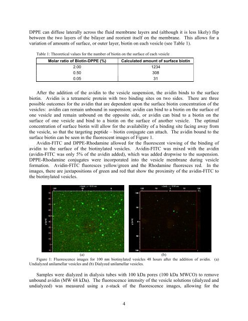
surface biot<strong>in</strong> can be seen <strong>in</strong> <strong>the</strong> fluorescent images <strong>of</strong> Figure 1.<br />
Avid<strong>in</strong>-FITC and DPPE-Rhodam<strong>in</strong>e allowed for <strong>the</strong> fluorescent view<strong>in</strong>g <strong>of</strong> <strong>the</strong> b<strong>in</strong>d<strong>in</strong>g <strong>of</strong><br />
avid<strong>in</strong> to <strong>the</strong> surface <strong>of</strong> <strong>the</strong> biot<strong>in</strong>ylated vesicles. Avid<strong>in</strong>-FITC was mixed with <strong>the</strong> avid<strong>in</strong><br />
(avid<strong>in</strong>-FITC was only 5% <strong>of</strong> <strong>the</strong> avid<strong>in</strong> added), which was added dropwise to <strong>the</strong> suspension.<br />
DPPE-Rhodam<strong>in</strong>e conjugates were <strong>in</strong>corporated <strong>in</strong>to <strong>the</strong> vesicle membrane dur<strong>in</strong>g vesicle<br />
formation. Avid<strong>in</strong>-FITC fluoresces yellow/green and <strong>the</strong> Rhodam<strong>in</strong>e fluoresces red. In <strong>the</strong><br />
images, <strong>the</strong>re are juxtapositions <strong>of</strong> green and red that show <strong>the</strong> proximity <strong>of</strong> <strong>the</strong> avid<strong>in</strong>-FITC to<br />
<strong>the</strong> biot<strong>in</strong>ylated vesicles.<br />
(a)<br />
(b)<br />
Figure 1: Fluorescence images for 100 nm biot<strong>in</strong>ylated vesicles 48 hours after <strong>the</strong> addition <strong>of</strong> avid<strong>in</strong>. (a)<br />
Undialyzed unilamellar vesicles and (b) Dialyzed unilamellar vesicles.<br />
Samples were dialyzed <strong>in</strong> dialysis tubes with 100 kDa pores (100 kDa MWCO) to remove<br />
unbound avid<strong>in</strong> (MW 68 kDa). <strong>The</strong> fluorescence <strong>in</strong>tensity <strong>of</strong> <strong>the</strong> vesicle solutions (dialyzed and<br />
undialyzed) was measured us<strong>in</strong>g a z-stack <strong>of</strong> <strong>the</strong> fluorescence images, allow<strong>in</strong>g for <strong>the</strong><br />
4