Simultaneous PET/MRI acquisition: Clinical potential in anatomically ...
Simultaneous PET/MRI acquisition: Clinical potential in anatomically ...
Simultaneous PET/MRI acquisition: Clinical potential in anatomically ...

You also want an ePaper? Increase the reach of your titles
YUMPU automatically turns print PDFs into web optimized ePapers that Google loves.
<strong>Simultaneous</strong> <strong>PET</strong>/<strong>MRI</strong> <strong>acquisition</strong>:<br />
<strong>Cl<strong>in</strong>ical</strong> <strong>potential</strong> <strong>in</strong> <strong>anatomically</strong><br />
focused and whole-body<br />
exam<strong>in</strong>ations<br />
Kathryn Fowler, MD, Jonathan McConathy, MD, PhD, Geetika Khanna, MD,<br />
Farrokh Dehdashti, MD, Tammie L.S. Benz<strong>in</strong>ger, MD, PhD, Michelle Miller-Thomas, MD,<br />
Matthew Parsons, MD, Constant<strong>in</strong>e Raptis, MD, Perry Grigsby, MD, Richard Laforest, PhD,<br />
Robert J. Gropler, MD, Vamsi Narra, MD, Barry A. Siegel, MD, John Kotyk, PhD,<br />
Agus Priatna, PhD, Robert McK<strong>in</strong>stry, MD, PhD, and Pamela K. Woodard, MD<br />
The cl<strong>in</strong>ical availability of hybrid<br />
positron emission tomography<br />
(<strong>PET</strong>) and magnetic resonance<br />
imag<strong>in</strong>g (<strong>MRI</strong>) systems offers medical<br />
imag<strong>in</strong>g a range of <strong>potential</strong> benefits <strong>in</strong><br />
oncologic, neurological, and cardiovascular<br />
applications. 1-5 One obvious<br />
benefit of simultaneous <strong>acquisition</strong> is<br />
improved image registration that allows<br />
optimal anatomic localization of<br />
<strong>PET</strong> f<strong>in</strong>d<strong>in</strong>gs. Other advantages <strong>in</strong>clude<br />
lower radiation dose <strong>in</strong> wholebody<br />
imag<strong>in</strong>g compared with <strong>PET</strong>/CT<br />
(computed tomography), shorter overall<br />
imag<strong>in</strong>g times, and the ability to simultaneously<br />
observe rapidly chang<strong>in</strong>g<br />
physiological and pathophysiological<br />
processes.<br />
Drs. Fowler, McConathy, Khanna,<br />
Dehdashti, Benz<strong>in</strong>ger, Miller-Thomas,<br />
Parsons, Raptis, Grigsby, Laforest,<br />
Gropler, Narra, Siegel, Kotyk, McK<strong>in</strong>stry,<br />
and Woodard practice at the Mall<strong>in</strong>ckrodt<br />
Institute of Radiology and the Alv<strong>in</strong><br />
J. Siteman Cancer Center, Wash<strong>in</strong>gton<br />
University School of Medic<strong>in</strong>e, St Louis,<br />
MO. Dr. Priatna is at R&D Collaborations,<br />
Siemens Healthcare, St Louis, MO.<br />
Most of the examples that follow are<br />
of patients scheduled for standard-ofcare<br />
cl<strong>in</strong>ical fluorodeoxyglucose (FDG)<br />
<strong>PET</strong>/CT who, with Institutional Review<br />
Board (IRB) approval, were recruited<br />
for, and consented to undergo, additional<br />
<strong>PET</strong>/MR imag<strong>in</strong>g. The simultaneous<br />
<strong>PET</strong>/<strong>MRI</strong> studies were acquired<br />
on a Biograph mMR system (Siemens<br />
Medical Systems, Erlangen, Germany)<br />
recently <strong>in</strong>stalled <strong>in</strong> the Center for <strong>Cl<strong>in</strong>ical</strong><br />
Imag<strong>in</strong>g Research (CCIR) at Wash<strong>in</strong>gton<br />
University School of Medic<strong>in</strong>e,<br />
St. Louis, MO. In this system, the <strong>PET</strong><br />
component of the scanner with MRcompatible<br />
avalanche photodiodes<br />
(APDs) is present with<strong>in</strong> the bore of a<br />
3.0 tesla (T) magnet equipped with total<br />
imag<strong>in</strong>g matrix and attenuation bodyarray<br />
and sp<strong>in</strong>e matrix coils.<br />
Unlike <strong>PET</strong>/CT, <strong>PET</strong>/<strong>MRI</strong> uses a soft<br />
tissue, segmentation attenuation-correction<br />
(AC) µ-map. The AC µ-map was<br />
generated utiliz<strong>in</strong>g a dual-echo VIBE<br />
Dixon sequence that separates water and<br />
fat with TE1/TE2 = 1.23 msec/2.46 msec,<br />
TR = 3.6 msec, left-right FOV = 500 mm<br />
and anterior-posterior FOV = 300 mm.<br />
June 2013<br />
www.appliedradiology.com APPLIED RADIOLOGY © n 9
SIMULTANEOUS <strong>PET</strong>/<strong>MRI</strong> ACQUISITION<br />
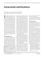
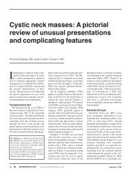
FIGURE 1. Left temporal lobe primitive neuroectodermal tumor (PNET), recurrent after resection<br />
and radiation to the bed. Arrows show focal region of FDG uptake, which was found to be<br />
recurrent tumor and radiation necrosis. A = T2 FLAIR, B = T1 MPRAGE precontrast, C = FDG-<br />
<strong>PET</strong>, D = TSE T2, E = T1 postcontrast, F = fused FDG-<strong>PET</strong> and T1 postcontrast images.<br />
The <strong>acquisition</strong>s were performed either<br />
<strong>in</strong> a s<strong>in</strong>gle station for dedicated<br />
<strong>PET</strong>/<strong>MRI</strong> or a multiple-station mode<br />
for whole-body imag<strong>in</strong>g as <strong>in</strong>dicated.<br />
Depend<strong>in</strong>g on the application, <strong>PET</strong><br />
images were simultaneously acquired<br />
with the anatomical sequences from<br />
<strong>MRI</strong>, such as HASTE for whole-body<br />
<strong>acquisition</strong>, MPRAGE for bra<strong>in</strong> imag<strong>in</strong>g,<br />
SPACE or HASTE for pelvic<br />
applications, or delayed, contrast-enhanced,<br />
T1-weighted, phase-sensitive<br />
<strong>in</strong>version-recovery imag<strong>in</strong>g for cardiac<br />
imag<strong>in</strong>g. Additional high-resolution<br />
<strong>MRI</strong> sequences were added for focused<br />
exam<strong>in</strong>ations; these <strong>in</strong>cluded<br />
high-resolution T2 TSE, diffusionweighted<br />
imag<strong>in</strong>g (DWI) or diffusion-tensor<br />
imag<strong>in</strong>g (DTI), and other<br />
sequences depend<strong>in</strong>g on the applications.<br />
Recent articles detail possible<br />
whole-body and dedicated protocols<br />
for oncologic imag<strong>in</strong>g with <strong>PET</strong>/MR;<br />
however, the optimal sequences for<br />
workflow efficiency and diagnostic<br />
yield are yet to be determ<strong>in</strong>ed <strong>in</strong> cl<strong>in</strong>ical<br />
practice. 6,7<br />
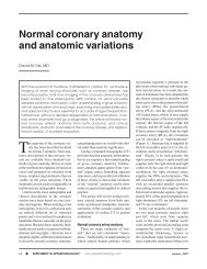
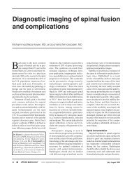
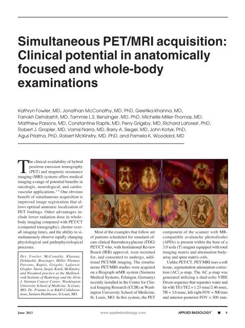
FIGURE 2. Amyloid imag<strong>in</strong>g with F-18 florbetapir. Panel A shows a normal control subject with no-to-sparse beta-amyloid plaques. Panel B<br />
shows a positive <strong>PET</strong>/<strong>MRI</strong> study, consistent with moderate to frequent beta-amyloid plaques.<br />
10 n APPLIED RADIOLOGY © www.appliedradiology.com June 2013
SIMULTANEOUS <strong>PET</strong>/<strong>MRI</strong> ACQUISITION<br />
<strong>Cl<strong>in</strong>ical</strong> cases<br />
The follow<strong>in</strong>g are some examples of<br />
the cl<strong>in</strong>ically relevant cases acquired for<br />
<strong>anatomically</strong> focused and whole-body<br />
exam<strong>in</strong>ations with the Biograph mMR.<br />
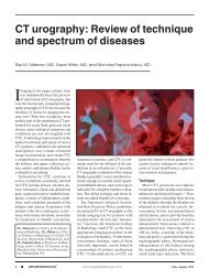
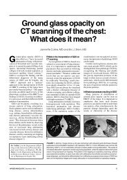
FIGURE 3. Buccal cancer. Images depict a metabolically active tumor <strong>in</strong> the right buccal<br />
space (arrows), which also is well shown on the DWI and ADC map to restrict diffusion. The<br />
patient had biopsy-proven metastatic <strong>in</strong>volvement of Ib lymph nodes with<strong>in</strong> the right submandibular<br />
space (open arrows).<br />
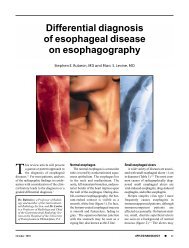
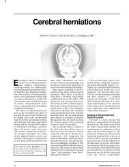
FIGURE 4. Multiple myeloma. Images depict a focus of metabolically active disease <strong>in</strong> the<br />
left humeral neck that is more conspicuous on <strong>MRI</strong> than on CT alone (arrows).<br />
Neuro-oncologic bra<strong>in</strong> imag<strong>in</strong>g<br />
While separately acquired bra<strong>in</strong> MR<br />
and FDG-<strong>PET</strong> images can be fused<br />
through various software tools, a comb<strong>in</strong>ed<br />
exam<strong>in</strong>ation permits streaml<strong>in</strong>ed<br />
patient care and improves diagnostic<br />
specificity. The convenience and reduced<br />
burden to the patient of a s<strong>in</strong>gle<br />
imag<strong>in</strong>g session is particularly relevant<br />
to patients with bra<strong>in</strong> tumors, who often<br />
have a reduced functional status.<br />
Figure 1 displays a panel of selected<br />
simultaneous FDG-<strong>PET</strong>/MR images of<br />
a 41-year-old man with a left temporal<br />
lobe primitive neuroectodermal tumor<br />
(PNET) treated with near-total resection,<br />
craniosp<strong>in</strong>al and resection-bed<br />
radiation therapy, and adjuvant chemotherapy.<br />
Follow-up <strong>MRI</strong> showed a<br />
grow<strong>in</strong>g, enhanc<strong>in</strong>g mass, and FDG-<br />
<strong>PET</strong>/<strong>MRI</strong> was performed to differentiate<br />
recurrent tumor from radiation<br />
necrosis. The white arrow denotes an<br />
enhanc<strong>in</strong>g nodule with markedly <strong>in</strong>creased<br />
FDG uptake, highly suspicious<br />
for recurrent PNET. Subsequent surgical<br />
resection demonstrated a mixture of<br />
recurrent PNET and radiation necrosis.<br />
Amyloid <strong>PET</strong> bra<strong>in</strong> imag<strong>in</strong>g<br />
The development of F-18-labeled<br />
<strong>PET</strong> tracers for imag<strong>in</strong>g beta-amyloid<br />
plaques that occur <strong>in</strong> Alzheimer’s disease<br />
(AD) has the <strong>potential</strong> to <strong>in</strong>crease<br />
the certa<strong>in</strong>ty of diagnosis <strong>in</strong> patients<br />
with cognitive impairment. One of<br />
these tracers, F-18 florbetapir, recently<br />
received FDA approval for cl<strong>in</strong>ical use,<br />
and several other amyloid <strong>PET</strong> tracers<br />
<strong>in</strong> late-phase cl<strong>in</strong>ical trials may soon<br />
become cl<strong>in</strong>ically available. If drugs<br />
that slow or reverse the development of<br />
AD dementia become cl<strong>in</strong>ically available,<br />
amyloid <strong>PET</strong> imag<strong>in</strong>g may be a<br />
key component of patient selection for<br />
treatment and for monitor<strong>in</strong>g response<br />
to therapy. The anatomic, volumetric,<br />
and functional data available through<br />
June 2013<br />
www.appliedradiology.com APPLIED RADIOLOGY © n 11
SIMULTANEOUS <strong>PET</strong>/<strong>MRI</strong> ACQUISITION<br />
relies primarily upon assessment of graywhite<br />
differentiation, with negative studies<br />
show<strong>in</strong>g higher activity <strong>in</strong> the white<br />
matter than <strong>in</strong> the cerebral cortex (Figure<br />
2) and positive studies show<strong>in</strong>g loss<br />
of gray-white contrast due to the tracer<br />
b<strong>in</strong>d<strong>in</strong>g to beta-amyloid plaques <strong>in</strong> the<br />
cerebral cortex (Figure 2).<br />
FIGURE 5. Normal myocardial uptake. <strong>Simultaneous</strong> <strong>acquisition</strong> of ECG-gated <strong>PET</strong> and<br />
delayed contrast-enhanced (DCE) cardiac MR images (simultaneous <strong>acquisition</strong> of MR<br />
2-po<strong>in</strong>t Dixon also acquired for attenuation correction prior to contrast <strong>in</strong>jection). <strong>PET</strong> data<br />
were acquired <strong>in</strong> list mode and b<strong>in</strong>ned. DCE-MR images acquired <strong>in</strong> diastole are fused with<br />
diastolic <strong>PET</strong> data to create the center image.<br />
FIGURE 6. Cervical cancer. Images (A) show a nodal metastasis <strong>in</strong> the left external-iliac<br />
cha<strong>in</strong> (arrows) <strong>in</strong> a patient with cervical cancer. Additional images (B) from a separate patient<br />
demonstrate T2-SPACE sequence, 3D isotropic <strong>acquisition</strong>, which allows reformatted images<br />
to be constructed <strong>in</strong> multiple planes without loss of resolution.<br />
<strong>MRI</strong> can complement the biochemical<br />
<strong>in</strong>formation obta<strong>in</strong>ed through amyloid<br />
<strong>PET</strong> and provide a comprehensive, s<strong>in</strong>gle-session<br />
neuroimag<strong>in</strong>g evaluation of<br />
patients with cognitive impairment.<br />
Figure 2 displays selected negative<br />
and positive F-18 florbetapir-<strong>PET</strong>/MR<br />
images from subjects enrolled <strong>in</strong> a research<br />
study. <strong>MRI</strong> provides excellent<br />
anatomic correlation for the localization<br />
of F-18 florbetapir accumulation <strong>in</strong> the<br />
white and gray matter and can also provide<br />
volumetric data for the detection of<br />
atrophy <strong>in</strong> regions like the hippocampus,<br />
which is often affected by AD. <strong>Cl<strong>in</strong>ical</strong><br />
<strong>in</strong>terpretation of F-18 florbetapir-<strong>PET</strong><br />
Head and neck oncology<br />
<strong>Simultaneous</strong> head and neck <strong>PET</strong>/<br />
<strong>MRI</strong> performs well compared with<br />
<strong>PET</strong>/CT <strong>in</strong> our <strong>in</strong>itial experience. In<br />
the head and neck, <strong>PET</strong>/<strong>MRI</strong> comb<strong>in</strong>es<br />
the metabolic <strong>in</strong>formation<br />
from FDG-<strong>PET</strong> with high spatial<br />
resolution, anatomic localization, and<br />
soft-tissue contrast from <strong>MRI</strong>. <strong>MRI</strong><br />
demonstrates better soft-tissue contrast<br />
than CT, permitt<strong>in</strong>g detection of<br />
per<strong>in</strong>eural spread. Software fusion of<br />
<strong>PET</strong> and <strong>MRI</strong> data is challeng<strong>in</strong>g <strong>in</strong><br />
the neck, and the simultaneous <strong>acquisition</strong><br />
of the <strong>MRI</strong> and <strong>PET</strong> data provides<br />
improved coregistration.<br />
Figure 3 illustrates images of a<br />
58-year-old man with a recent diagnosis<br />
of squamous cell carc<strong>in</strong>oma of the right<br />
buccal space and metastatic <strong>in</strong>volvement<br />
of right submandibular lymph<br />
nodes. Contrast-enhanced <strong>MRI</strong> shows<br />
excellent anatomic detail, and diffusion-weighted<br />
images demonstrate high<br />
contrast between the lymph nodes and<br />
adjacent fat <strong>in</strong> the submandibular space,<br />
which correlate to the areas of <strong>in</strong>creased<br />
FDG uptake.<br />
Multiple myeloma and<br />
bone imag<strong>in</strong>g<br />
The comb<strong>in</strong>ation of functional and<br />
morphologic <strong>MRI</strong> sequences with <strong>PET</strong><br />
imag<strong>in</strong>g is of <strong>potential</strong> value <strong>in</strong> the<br />
evaluation of osseous metastases and<br />
primary bone neoplasms, such as multiple<br />
myeloma. DWI, T2 fat-suppressed<br />
sequences, and T1-TSE images can all<br />
depict bone lesions that may not be evident<br />
on CT or conventional bone sc<strong>in</strong>tigraphy<br />
alone. While the relationship<br />
of diffusion restriction with disease<br />
status is complex, ADC values and appearance<br />
on DWI, <strong>in</strong> conjunction with<br />
changes <strong>in</strong> FDG uptake, may be useful<br />
12 n APPLIED RADIOLOGY © www.appliedradiology.com June 2013
SIMULTANEOUS <strong>PET</strong>/<strong>MRI</strong> ACQUISITION<br />
FIGURE 7. B-cell lymphoma undergo<strong>in</strong>g restag<strong>in</strong>g after chemotherapy. Anterior <strong>PET</strong>/<strong>MRI</strong> (A) and <strong>PET</strong>/CT (B) maximal <strong>in</strong>tensity projection<br />
(MIP) images show a hypermetabolic focus <strong>in</strong> the mediast<strong>in</strong>um (arrow), which was pathologically proven recurrent disease. A hypermetabolic<br />
lymph node (open arrow) is also suspicious for disease <strong>in</strong>volvement. A third lesion <strong>in</strong> the left kidney (solid arrowhead) is difficult to differentiate<br />
from collect<strong>in</strong>g system activity on the CT (F); however, axial HASTE (C), fused (D), and diffusion-weighted (E) MR images show that this represents<br />
a renal lesion.<br />
for monitor<strong>in</strong>g response to therapy <strong>in</strong><br />
neoplasms <strong>in</strong>volv<strong>in</strong>g the bone.<br />
Figure 4 consists of images of a<br />
62-year-old woman with biopsy-proven<br />
multiple myeloma present<strong>in</strong>g for <strong>in</strong>itial<br />
stag<strong>in</strong>g. A large, left humeral neck lesion<br />
is clearly depicted on DWI, ADC, and<br />
T2-<strong>in</strong>version recovery, fat-suppressed<br />
MR sequences, with <strong>in</strong>creased FDG uptake<br />
shown on the fused image. This lesion<br />
is less conspicuous on CT alone.<br />
Cardiac imag<strong>in</strong>g<br />
Cardiac <strong>PET</strong>/<strong>MRI</strong> could play a role<br />
<strong>in</strong> both cardiac ischemia and cardiac<br />
viability assessment. Potential cl<strong>in</strong>ical<br />
protocols, play<strong>in</strong>g on the strengths<br />
of both modalities, <strong>in</strong>clude stable chest<br />
pa<strong>in</strong> assessment, perform<strong>in</strong>g c<strong>in</strong>e <strong>MRI</strong><br />
cardiac function assessment, N-13<br />
ammonia or Rb-82 <strong>PET</strong> myocardial<br />
perfusion imag<strong>in</strong>g, and delayed contrast-enhanced<br />
<strong>in</strong>version-recovery <strong>in</strong>farct<br />
imag<strong>in</strong>g <strong>in</strong> a s<strong>in</strong>gle exam<strong>in</strong>ation.<br />
Comb<strong>in</strong>ed FDG and delayed contrastenhanced<br />
<strong>in</strong>version-recovery cardiac<br />
imag<strong>in</strong>g permit co-localized, simultaneously<br />
acquired functional and anatomic<br />
viability assessment that, theoretically,<br />
could play a role <strong>in</strong> imag<strong>in</strong>g-directed<br />
ventricular tachycardia radiofrequency<br />
ablation or direct biventricular pac<strong>in</strong>g <strong>in</strong><br />
dyssynchrony.<br />
Figure 5 shows simultaneously acquired<br />
ECG-gated <strong>PET</strong> and delayed<br />
contrast-enhanced (DCE) cardiac MR<br />
images (simultaneous <strong>acquisition</strong> of<br />
MR 2-po<strong>in</strong>t Dixon also acquired for<br />
AC), allow<strong>in</strong>g for precise fusion imag<strong>in</strong>g.<br />
<strong>PET</strong> data were acquired <strong>in</strong> list<br />
mode, b<strong>in</strong>ned, and reconstructed <strong>in</strong>to 3<br />
phases. DCE MR images acquired <strong>in</strong> diastole<br />
are fused with diastolic <strong>PET</strong> data<br />
to create the center image. This patient<br />
has a normal heart.<br />
Cervical and pelvic cancers<br />
FDG-<strong>PET</strong>/CT and <strong>MRI</strong> are well<br />
established for stag<strong>in</strong>g and monitor<strong>in</strong>g<br />
treatment response <strong>in</strong> patients with<br />
cervical cancer and other pelvic malignancies.<br />
<strong>PET</strong> data provide tumor<br />
volume estimation, allow<strong>in</strong>g for accurate<br />
radiation therapy plann<strong>in</strong>g, as<br />
well as prognostic <strong>in</strong>formation related<br />
to progression-free survival. 4-7 Highresolution<br />
<strong>MRI</strong> provides exquisite<br />
soft-tissue detail for local stag<strong>in</strong>g and<br />
presurgical plann<strong>in</strong>g. The comb<strong>in</strong>ation<br />
of metabolic <strong>in</strong>formation derived from<br />
FDG-<strong>PET</strong> with high-resolution <strong>MRI</strong> of<br />
the pelvis shows promise <strong>in</strong> both cl<strong>in</strong>ical<br />
management and <strong>potential</strong> research<br />
June 2013<br />
www.appliedradiology.com APPLIED RADIOLOGY © n 13
SIMULTANEOUS <strong>PET</strong>/<strong>MRI</strong> ACQUISITION<br />
opportunities <strong>in</strong> correlat<strong>in</strong>g functional<br />
<strong>MRI</strong> with tumor metabolism.<br />
Given the complex anatomy of the<br />
pelvis, multiplanar and high-resolution<br />
T2 TSE are ma<strong>in</strong>stay sequences. Us<strong>in</strong>g<br />
a 3-dimensional (3D) isotropic dataset,<br />
such as T2 SPACE, a s<strong>in</strong>gle <strong>acquisition</strong><br />
can yield high-resolution images with<br />
the option of <strong>in</strong>f<strong>in</strong>ite reformatt<strong>in</strong>g without<br />
loss of resolution. DWI provides<br />
improved conspicuity of lymph nodes,<br />
and ADC values for primary cervical<br />
malignancy have been correlated with<br />
standardized uptake values (SUVs) for<br />
FDG, permitt<strong>in</strong>g an additional non<strong>in</strong>vasive<br />
biomarker of disease response.<br />
Additionally, volumetric-<strong>in</strong>terpolated<br />
breath-held exam<strong>in</strong>ation (VIBE) can<br />
provide excellent anatomic detail of<br />
pelvic structures. Radial VIBE allows<br />
for a free-breath<strong>in</strong>g <strong>acquisition</strong> with<br />
acceptable resolution and no motion<br />
artifact.<br />
Figure 6 consists of images of a<br />
58-year-old woman with newly diagnosed<br />
cervical cancer present<strong>in</strong>g for<br />
<strong>in</strong>itial stag<strong>in</strong>g. Additional images from<br />
a separate patient who underwent total<br />
abdom<strong>in</strong>al hysterectomy and bilateral<br />
salp<strong>in</strong>go-oophorectomy demonstrate<br />
the utility of T2-SPACE multiplanar<br />
reformats.<br />
Pediatric imag<strong>in</strong>g<br />
<strong>PET</strong>/<strong>MRI</strong> is especially appeal<strong>in</strong>g<br />
<strong>in</strong> the pediatric population, as it is associated<br />
with less radiation exposure<br />
than <strong>PET</strong>/CT without compromis<strong>in</strong>g<br />
anatomic image quality. A recent study<br />
performed at the University of Leipzig<br />
<strong>in</strong> Germany showed that the effective<br />
dose of a <strong>PET</strong>/<strong>MRI</strong> scan is only about<br />
20% that of the equivalent <strong>PET</strong>/CT<br />
exam<strong>in</strong>ation. Children with systemic<br />
malignancies rout<strong>in</strong>ely evaluated with<br />
<strong>PET</strong>/CT (such as lymphoma) could<br />
benefit from the reduced radiation exposure<br />
of <strong>PET</strong>/<strong>MRI</strong>.<br />
<strong>Simultaneous</strong>ly acquir<strong>in</strong>g <strong>PET</strong> and<br />
<strong>MRI</strong> data comb<strong>in</strong>es the advantages<br />
of two previously separate advancedimag<strong>in</strong>g<br />
modalities, a tremendous advantage<br />
<strong>in</strong> young children requir<strong>in</strong>g<br />
sedation/anesthesia for imag<strong>in</strong>g. The<br />
improved soft-tissue contrast and molecular<br />
imag<strong>in</strong>g (such as diffusion and<br />
perfusion imag<strong>in</strong>g) abilities of <strong>MRI</strong><br />
permit better detection of lymph-node<br />
and visceral disease than does CT. <strong>PET</strong>/<br />
<strong>MRI</strong> also holds great <strong>potential</strong> <strong>in</strong> evaluat<strong>in</strong>g<br />
soft-tissue malignancies, such as<br />
sarcomas, <strong>in</strong> the pediatric population.<br />
Figure 7 presents the images of a<br />
16-year-old boy with diffuse, large B-<br />
cell lymphoma undergo<strong>in</strong>g restag<strong>in</strong>g<br />
after chemotherapy. Excellent <strong>PET</strong> and<br />
<strong>MRI</strong> coregistration helps separate the<br />
renal lesion (solid arrowhead) from excreted<br />
FDG <strong>in</strong> the collect<strong>in</strong>g system.<br />
Conclusion<br />
<strong>PET</strong>/<strong>MRI</strong> shows promise for multiple<br />
cl<strong>in</strong>ical applications through the<br />
comb<strong>in</strong>ation of the improved soft-tissue<br />
contrast of <strong>MRI</strong> with lower radiation<br />
dose, and the <strong>potential</strong> for better correlation<br />
of <strong>PET</strong> f<strong>in</strong>d<strong>in</strong>gs to anatomy given<br />
the simultaneous <strong>acquisition</strong>. Several<br />
challenges are evident <strong>in</strong> develop<strong>in</strong>g<br />
optimal protocols, <strong>in</strong>clud<strong>in</strong>g optimal<br />
MR-sequence parameters, motion correction,<br />
and validation of the quantitative<br />
accuracy of <strong>PET</strong> with <strong>MRI</strong>-based<br />
attenuation correction. In some cases,<br />
the comb<strong>in</strong>ation of SUVs measured on<br />
<strong>PET</strong> and ADC values measured on diffusion-weighted<br />
<strong>MRI</strong> may prove more<br />
specific than subjective assessment<br />
alone <strong>in</strong> differentiat<strong>in</strong>g tumor from<br />
surround<strong>in</strong>g tissue. The <strong>potential</strong> for<br />
benefit from <strong>PET</strong>/<strong>MRI</strong> <strong>acquisition</strong> also<br />
exists <strong>in</strong> receptor-targeted oncologic<br />
imag<strong>in</strong>g, dementia assessment, and cardiac<br />
and atherosclerosis imag<strong>in</strong>g.<br />
Acknowledgements<br />
We acknowledge the <strong>in</strong>valuable assistance<br />
of Jennifer Frye, Glenn Foster,<br />
L<strong>in</strong>da Becker, Debra Hew<strong>in</strong>g, Michael<br />
Harrod, Tim Street, and Betsy Thomas.<br />
References<br />
1. Pichler BJ, Judenhofer MS, Wehrl HF. <strong>PET</strong>/<strong>MRI</strong><br />
hybrid imag<strong>in</strong>g: Devices and <strong>in</strong>itial results. Eur<br />
Radiol. 2008;18:1077-1786.<br />
2. Antoch G, Bockisch A. Comb<strong>in</strong>ed <strong>PET</strong>/<strong>MRI</strong>: A<br />
new dimension <strong>in</strong> whole-body oncology imag<strong>in</strong>g?<br />
Eur J Nucl Med Mol Imag<strong>in</strong>g. 2009;36 Suppl<br />
1:S113-20.<br />
3. Wehrl HF, Sauter AW, Judenhofer MS, Pichler BJ.<br />
Comb<strong>in</strong>ed <strong>PET</strong>/MR imag<strong>in</strong>g--technology and applications.<br />
Technol Cancer Res Treat. 2010;9:5-20.<br />
4. Buchbender C, Heusner TA, Lauenste<strong>in</strong> TC,<br />
et al. Oncologic <strong>PET</strong>/<strong>MRI</strong>, Part 1: Tumors of the<br />
bra<strong>in</strong>, head and neck, chest, abdomen, and pelvis.<br />
J Nucl Med. 2012;53:928-938.<br />
5. Buchbender C, Heusner TA, Lauenste<strong>in</strong> TC, et<br />
al. Oncologic <strong>PET</strong>/<strong>MRI</strong>, Part 2: Bone tumors, softtissue<br />
tumors, melanoma, lymphoma. J Nucl Med.<br />
2012;53:1244-1252.<br />
6. Drzezga A, Souvatzoglou M, Eiber M, et al.<br />
First cl<strong>in</strong>ical experience with <strong>in</strong>tegrated wholebody<br />
<strong>PET</strong>/MR: Comparison to <strong>PET</strong>/CT <strong>in</strong> patients<br />
with oncologic diagnoses. J Nucl Med. 2012;53:<br />
845-855.<br />
7. Mart<strong>in</strong>ez-Moller A, Eiber M, Nekolla S, et al.<br />
Workflow and scan protocol considerations for<br />
<strong>in</strong>tegrated whole-body <strong>PET</strong>/<strong>MRI</strong> <strong>in</strong> oncology. J<br />
Nucl Med. 2012;53:1415-1426.<br />
8. Kidd EA, Siegel BA, Dehdashti F, Grigsby PW.<br />
The standardized uptake value for F-18 fluorodeoxyglucose<br />
is a sensitive predictive biomarker for<br />
cervical cancer treatment response and survival.<br />
Cancer. 2007;110:1738-1744.<br />
9. Kidd EA, Grigsby PW. Intratumoral metabolic<br />
heterogeneity of cervical cancer. Cl<strong>in</strong> Cancer Res.<br />
2008; 14:5236-5241.<br />
10. Schwarz JK, Siegel BA, Dehdashti F, Grigsby<br />
PW. Association of post-therapy positron emission<br />
tomography with tumor response and survival <strong>in</strong><br />
cervical carc<strong>in</strong>oma. JAMA. 2007;298:2289-2295.<br />
11. Olsen JR, Esthappan J, Dewees T, et al.<br />
Tumor volume and subvolume concordance<br />
between FDG-<strong>PET</strong>/CT and diffusion-weighted<br />
<strong>MRI</strong> for squamous cell carc<strong>in</strong>oma of the cervix. J<br />
Magn Reson Imag<strong>in</strong>g. 2013; 37:431-434.<br />
12. Hirsch FW, Sattler B, Sorge I, et al. <strong>PET</strong>/MR<br />
<strong>in</strong> children. Initial cl<strong>in</strong>ical experience <strong>in</strong> paediatric<br />
oncology us<strong>in</strong>g an <strong>in</strong>tegrated <strong>PET</strong>/MR scanner.<br />
Pediatr Radiol. 2013 Jan 11. [Epub ahead of pr<strong>in</strong>t].<br />
14 n APPLIED RADIOLOGY © www.appliedradiology.com June 2013