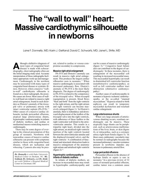
The “wall to wall” heart: Massive cardiothymic silhouette in newborns
The “wall to wall” heart: Massive cardiothymic silhouette in newborns
The “wall to wall” heart: Massive cardiothymic silhouette in newborns
Create successful ePaper yourself
Turn your PDF publications into a flip-book with our unique Google optimized e-Paper software.
<strong>The</strong> <strong>“wall</strong> <strong>to</strong> <strong>wall”</strong> <strong>heart</strong>:<br />
<strong>Massive</strong> <strong>cardiothymic</strong> <strong>silhouette</strong><br />
<strong>in</strong> <strong>newborns</strong><br />
A lthough<br />
Lane F. Donnelly, MD; Kar<strong>in</strong> J. Gelfand; David C. Schwartz, MD; Janet L. Strife, MD<br />
a def<strong>in</strong>itive diagnosis of<br />
most types of congenital <strong>heart</strong><br />
disease is made with echocardiography,<br />
chest radiographs often are<br />
the <strong>in</strong>itial imag<strong>in</strong>g study used. Accurate<br />
<strong>in</strong>terpretation of these radiographs facilitates<br />
appropriate work-up and management.<br />
Cardiomegaly <strong>in</strong> the newborn<br />
may be associated with many types of<br />
congenital <strong>heart</strong> disease or systemic illness.<br />
However, when a massive <strong>“wall</strong><strong>to</strong>-<strong>wall”</strong><br />
<strong>cardiothymic</strong> <strong>silhouette</strong> is<br />
found on a chest radiograph, the possible<br />
causes are fewer. Most cases of wall<strong>to</strong>-wall<br />
<strong>heart</strong> are related <strong>to</strong> massive right<br />
atrial enlargement, found <strong>in</strong> such disorders<br />
as Ebste<strong>in</strong>’s anomaly of the tricuspid<br />
valve or pulmonary atresia with<br />
<strong>in</strong>tact ventricular septum (PA-IVS). 1-3<br />
Other causes of a massive cardiac <strong>silhouette</strong><br />
<strong>in</strong>clude pericardial effusions,<br />
atypical large arteriovenous shunts,<br />
hypertrophic cardiomyopathy <strong>in</strong> <strong>in</strong>fants<br />
of diabetic mothers, and cardiac or<br />
mediast<strong>in</strong>al masses mimick<strong>in</strong>g massive<br />
cardiomegaly.<br />
<strong>The</strong>re may be some degree of overlap<br />
<strong>in</strong> the cl<strong>in</strong>ical presentation of this<br />
group of neonates. Newborns with<br />
noncardiac causes of massive cardiac<br />
<strong>silhouette</strong> can present with symp<strong>to</strong>ms<br />
similar <strong>to</strong> those with congenital <strong>heart</strong><br />
disease, which are tachypnea and<br />
wheez<strong>in</strong>g. Cyanosis also may be pres-<br />
Dr. Donnelly is with Duke University<br />
Medical Center <strong>in</strong> Durham, NC. Ms.<br />
Gelfand, Dr. Schwartz, and Dr. Strife<br />
are with Children’s Hospital Medical<br />
Center <strong>in</strong> C<strong>in</strong>c<strong>in</strong>nati, OH.<br />
APPLIED RADIOLOGY, December 1997<br />
ent, related <strong>to</strong> cardiac or venous compromise<br />
secondary <strong>to</strong> compression.<br />
<strong>Massive</strong> right atrial enlargement<br />
PA-IVS and Ebste<strong>in</strong>’s anomaly can<br />
result <strong>in</strong> massive right atrial enlargement.<br />
This produces the largest cardiac<br />
<strong>silhouette</strong>s seen <strong>in</strong> neonates. 1-3 When<br />
massive cardiomegaly is associated with<br />
decreased pulmonary flow, Ebste<strong>in</strong>’s<br />
anomaly or PA-IVS is the most likely<br />
diagnosis. <strong>The</strong> degree of cardiomegaly<br />
<strong>in</strong> PA-IVS is related <strong>to</strong> the competency<br />
of the tricuspid valve. 1 When tricuspid<br />
regurgitation is present, blood flows<br />
“back and forth” from the right ventricle<br />
<strong>to</strong> the right atrium, and the right atrium<br />
and right ventricle can become massively<br />
enlarged (figure 1). 1 In Ebste<strong>in</strong>’s<br />
anomaly, there is redundancy and downward<br />
displacement of the leaflets of the<br />
tricuspid valve <strong>in</strong><strong>to</strong> the right ventricle,<br />
with adherence of these leaflets <strong>to</strong> the<br />
right ventricular wall distal <strong>to</strong> the atrioventricular<br />
annulus. This results <strong>in</strong> a<br />
tripartite right <strong>heart</strong>, divided <strong>in</strong><strong>to</strong> the<br />
right atrium, an atrialized right ventricle<br />
(portion of the right ventricle located<br />
between the atrioventricular annulus and<br />
the displaced tricuspid valve), and the<br />
right ventricle. 4 A comb<strong>in</strong>ation of functional<br />
and structural obstruction leads <strong>to</strong><br />
massive enlargement of the ana<strong>to</strong>mic<br />
right atrium and atrialized right ventricle<br />
(figure 2). 3,4<br />
Hypertrophic cardiomyopathy <strong>in</strong><br />
<strong>in</strong>fants of diabetic mothers<br />
In <strong>in</strong>fants of diabetic mothers, an<br />
<strong>in</strong>crease <strong>in</strong> myocardial mass may result<br />
<strong>in</strong> hypertrophic cardiomyopathy and<br />
can be a cause of massive cardiomegaly<br />
(figure 3). 5,6 Congestive <strong>heart</strong> failure<br />
also can contribute <strong>to</strong> the degree of cardiomegaly.<br />
5 In these neonates, there is<br />
enlargement of the myocardial cell<br />
result<strong>in</strong>g <strong>in</strong> <strong>in</strong>creased myocardial mass.<br />
This myocardial hypertrophy can result<br />
<strong>in</strong> dim<strong>in</strong>ished left ventricular function<br />
(nonobstructive cardiomyopathy), as<br />
well as left ventricular outflow tract<br />
obstruction (obstructive cardiomyopathy).<br />
5<br />
Another cause of cardiomyopathy <strong>in</strong><br />
neonates is hypoxic-ischemic cardiomyopathy,<br />
or the so-called “stunned<br />
myocardium.” Hypoxia related <strong>to</strong> birth<br />
asphyxia can result <strong>in</strong> temporary<br />
myocardial dysfunction and resultant<br />
cardiac enlargement.<br />
Large arteriovenous shunts<br />
When very large amounts of arteriovenous<br />
shunt<strong>in</strong>g occurs, resultant cardiomegaly<br />
can be massive. <strong>The</strong>se<br />
atypically large shunts can be peripheral,<br />
such as arteriovenous malformations<br />
of the liver, bra<strong>in</strong>, or sk<strong>in</strong> (figure<br />
4), or <strong>in</strong>tracardiac, such as a coronary<br />
artery-right <strong>heart</strong> fistula (figure 5). 7 In<br />
these cases, pulmonary vascularity may<br />
be normal on radiographs because the<br />
<strong>in</strong>creased pulmonary vascular resistance<br />
present <strong>in</strong> <strong>newborns</strong> impedes any<br />
<strong>in</strong>crease <strong>in</strong> pulmonary arterial flow.<br />
Large pericardial effusions<br />
Pericardial effusion <strong>in</strong> the absence of<br />
hydrops fetalis or sepsis is rare <strong>in</strong><br />
neonates. 8 In a previous review of 32<br />
cases of neonatal pericardial effusion,<br />
cardiac tumor was a common cause<br />
23
A C<br />
B<br />
FIGURE 1. Pulmonary atresia with <strong>in</strong>tact<br />
<strong>in</strong>traventricular septum associated with tricuspid<br />
<strong>in</strong>sufficiency. (A) Chest radiograph<br />
shows massive cardiomegaly with a <strong>“wall</strong><strong>to</strong>-wall<br />
<strong>heart</strong>.” (B) Right <strong>heart</strong> with contrast<br />
<strong>in</strong>jection shows massive dilatation of the<br />
right atrium (RA) and right ventricle (RV).<br />
(C) Left <strong>heart</strong> with contrast <strong>in</strong>jection is normal<br />
sized, with a massively dilated right<br />
atrium and ventricle constitut<strong>in</strong>g the bulk of<br />
the cardiac shadow. (D) Au<strong>to</strong>psy specimen<br />
shows massive enlargement of the right<br />
atrium (arrows).<br />
A<br />
D<br />
FIGURE 2. Ebste<strong>in</strong>’s anomaly. Chest radiograph<br />
shows massive cardiomegaly with<br />
umbilical venous catheter (arrow) curled<br />
with<strong>in</strong> the dilated right atrium. Pulmonary<br />
vascularity is decreased.<br />
FIGURE 3. Hypertrophic cardiomyopathy <strong>in</strong><br />
an <strong>in</strong>fant of a diabetic mother. Radiograph<br />
shows marked cardiomegaly and prom<strong>in</strong>ent,<br />
<strong>in</strong>dist<strong>in</strong>ct pulmonary vasculature consistent<br />
with pulmonary venous congestion.<br />
FIGURE 4. Large cavernous hemangioma<br />
of the scalp and upper trunk. (A) Pho<strong>to</strong>graph<br />
of an <strong>in</strong>fant with cavernous hemangioma<br />
(arrow). (B) Frontal radiograph<br />
shows marked cardiomegaly and normal<br />
pulmonary vascular mark<strong>in</strong>gs. <strong>The</strong> great<br />
arteries and ve<strong>in</strong>s are enlarged secondary<br />
<strong>to</strong> <strong>in</strong>creased flow <strong>to</strong> the head, widen<strong>in</strong>g the<br />
superior mediast<strong>in</strong>um.<br />
24 APPLIED RADIOLOGY, December 1997<br />
B
A B C<br />
FIGURE 5. Coronary artery <strong>to</strong> right atrial fistula. (A) Frontal radiograph shows massive cardiomegaly and prom<strong>in</strong>ent and <strong>in</strong>dist<strong>in</strong>ct<br />
pulmonary vasculature. (B) Axial T1-weighted MR image shows dilated left coronary artery (arrow) from left coronary artery<br />
<strong>to</strong> right atrial fistula. (RA= right atrium; P = pulmonary artery; A = descend<strong>in</strong>g aorta.) (C) Coronal T1-weighted MR image shows<br />
marked dilatation of the right atrium [RA] constitut<strong>in</strong>g the majority of the enlarged cardiac <strong>silhouette</strong>.<br />
A B C<br />
FIGURE 6. Pericardial effusion secondary <strong>to</strong> left atrial hemangioendothelioma. (A) Initial radiograph shows enlarged cardiac <strong>silhouette</strong><br />
and a hemivertebrae. (B) Gadol<strong>in</strong>ium-enhanced, axial T1-weighted MRI shows a well-def<strong>in</strong>ed enhanc<strong>in</strong>g mass (arrow)<br />
<strong>in</strong>volv<strong>in</strong>g the lateral wall of the left atrium. <strong>The</strong>re is low signal <strong>in</strong> the pericardial sac consistent with residual air post-pericardialcentesis<br />
(small arrows). (C) After dra<strong>in</strong>age of the pericardial fluid, the cardiac <strong>silhouette</strong> is normal <strong>in</strong> size.<br />
A B<br />
A B<br />
APPLIED RADIOLOGY, December 1997<br />
FIGURE 7. Cardiac rhabdomyoma <strong>in</strong> a newborn<br />
with tuberous sclerosis. (A) Frontal<br />
radiograph shows massive cardiomegaly<br />
mimick<strong>in</strong>g Ebste<strong>in</strong>’s anomaly. (B) Substernal<br />
longitud<strong>in</strong>al sonogram demonstrates a<br />
large <strong>in</strong>filtrative mass (large arrows) aris<strong>in</strong>g<br />
from and surround<strong>in</strong>g the <strong>heart</strong> (small<br />
arrows).<br />
FIGURE 8. Mediast<strong>in</strong>al tera<strong>to</strong>ma. (A)<br />
Frontal radiograph shows massive cardiomegaly.<br />
(B) Angiogram shows displacement<br />
of both opacified right and left cardiac<br />
chambers secondary <strong>to</strong> a large left-sided<br />
mediast<strong>in</strong>al mass (arrows).<br />
27
A B<br />
A<br />
FIGURE 10. Apparent cardiomegaly secondary<br />
<strong>to</strong> technical fac<strong>to</strong>rs. (A) Portable,<br />
frontal anterior-posterior radiograph with lordotic<br />
position<strong>in</strong>g demonstrates apparent cardiac<br />
enlargement. (B) Lateral chest<br />
radiograph shows normal <strong>heart</strong> size. <strong>The</strong><br />
apparent cardiomegaly seen on the frontal<br />
radiograph is secondary <strong>to</strong> technical fac<strong>to</strong>rs.<br />
(38%), with tera<strong>to</strong>ma and cavernous<br />
hemangioma/hemangioma be<strong>in</strong>g the<br />
most common types. 8 Other common<br />
causes <strong>in</strong>cluded thyroid dysfunction<br />
(21%), <strong>in</strong>fection (12%), and diaphragmatic<br />
hernia <strong>in</strong><strong>to</strong> the pericardial sac<br />
(16%). 8 When large enough, pericardial<br />
effusions can cause massive enlargement<br />
of the cardiac <strong>silhouette</strong> (figure 6).<br />
Masses mimick<strong>in</strong>g cardiac chamber<br />
enlargement<br />
Chest masses can sometimes produce<br />
the radiographic <strong>“wall</strong>-<strong>to</strong>-<strong>wall”</strong> appearance<br />
of the <strong>heart</strong> <strong>in</strong> <strong>newborns</strong>, mimick<strong>in</strong>g<br />
diseases such as Ebste<strong>in</strong>’s anomaly.<br />
We have seen this occur with cardiac<br />
masses such as rhabdomyomas (figure<br />
B<br />
7), mediast<strong>in</strong>al masses such as tera<strong>to</strong>mas<br />
(figure 8), and congenital<br />
diaphragmatic hernias on early films,<br />
before gas enters the bowel (figure 9).<br />
Children with tuberous sclerosis are predisposed<br />
<strong>to</strong> develop<strong>in</strong>g rhabdomyomas.<br />
Technical fac<strong>to</strong>rs<br />
Many technical fac<strong>to</strong>rs can <strong>in</strong>fluence<br />
the apparent cardiac size seen on frontal<br />
radiographs. In neonatal <strong>in</strong>tensive care<br />
units, radiographs often are obta<strong>in</strong>ed<br />
us<strong>in</strong>g the portable anterior-posterior<br />
technique <strong>in</strong> frontal projection only.<br />
Anterior-posterior projection, lordotic<br />
or rotated position<strong>in</strong>g, and large focusfilm<br />
distance all may cause the magnification<br />
of apparent cardiac size. 9 Under<br />
FIGURE 9. Congenital diaphragmatic hernia.<br />
(A) Initial radiograph after birth shows<br />
what appears <strong>to</strong> be massive cardiomegaly.<br />
Note <strong>in</strong>dist<strong>in</strong>ctness of the left hemidiaphragm.<br />
(B) Delayed frontal radiograph<br />
shows aeration of the bowel with<strong>in</strong> the left<br />
hemithorax consistent with a congenital<br />
diaphragmatic hernia. (Image courtesy of<br />
Charles A. Good<strong>in</strong>g, MD, University of California,<br />
San Francisco, CA.)<br />
these circumstances, a normal <strong>heart</strong> can<br />
appear <strong>to</strong> be enlarged (figure 10), especially<br />
if the film is obta<strong>in</strong>ed dur<strong>in</strong>g<br />
expiration. Additionally, a normal thymus<br />
can appear large relative <strong>to</strong> the<br />
newborn mediast<strong>in</strong>um and mimic cardiomegaly<br />
on frontal radiographs. In<br />
cases where cardiac enlargement is <strong>in</strong><br />
question on frontal radiographs, lateral<br />
radiographs are helpful <strong>in</strong> determ<strong>in</strong><strong>in</strong>g<br />
whether cardiac enlargement is true or<br />
artifactual. AR<br />
REFERENCES<br />
1. Kanjuh VI, Stevenson JE, Amplatz K, et al:<br />
Congenitally unguarded tricuspid orifice with coexistent<br />
pulmonary atresia. Circulation 30:911-<br />
915, 1964.<br />
2. Anderson KR, Lie JT: Pathologic ana<strong>to</strong>my of<br />
Ebste<strong>in</strong>’s anomaly of the <strong>heart</strong> revisited. Am J Cardiol<br />
41:739-745, 1978.<br />
3. Strife JL, Bisset GS III: Cardiovascular system.<br />
In: Kirks DR (ed), Practical Pediatric Imag<strong>in</strong>g, pp<br />
417-513. Bos<strong>to</strong>n, Little, Brown and Company,<br />
1991.<br />
4. Bialos<strong>to</strong>zky D, Horwitz S, Esp<strong>in</strong>o-Vela J:<br />
Ebste<strong>in</strong>’s malformation of the tricuspid valve: A<br />
review of 65 cases. Am J Cardiol 29:826-836,<br />
1972.<br />
5. Dunn V, Nixon GW, Jaffe RB, Condon VR:<br />
Infants of diabetic mothers: Radiographic manifestations.<br />
AJR 137:123-128, 1981.<br />
6. Way GL, Wolfe RR, Eshaghpour E, et al: <strong>The</strong><br />
natural his<strong>to</strong>ry of hypertrophic cardiomyopathy <strong>in</strong><br />
<strong>in</strong>fants of diabetic mothers. J Pediatrics 95:1020-<br />
1025, 1979.<br />
7. Jaffe RB, Glancy DL, Epste<strong>in</strong> SE, et al: Coronary<br />
arterial-right <strong>heart</strong> fistulae: Long-term observations<br />
<strong>in</strong> seven patients. Circulation 47:133-143,<br />
1973.<br />
8. Cartagena AM, Lev<strong>in</strong> TL, Issenberg H, Goldman<br />
HS: Pericardial effusion and cardiac hemangioma<br />
<strong>in</strong> the neonate. Pediatr Radiol 23:384-385,<br />
1993.<br />
9. Amplatz K, Moller JH: Radiology of Congenital<br />
Heart Disease, pp 55-113. St. Louis, Mosby-Year<br />
Book, Inc., 1993.<br />
28 APPLIED RADIOLOGY, December 1997


