Download PDF - Applied Radiology Online
Download PDF - Applied Radiology Online
Download PDF - Applied Radiology Online

You also want an ePaper? Increase the reach of your titles
YUMPU automatically turns print PDFs into web optimized ePapers that Google loves.
Pulmonary embolism (PE) and deep venous thrombosis (DVT)<br />
are components of the same disease entity, accounting for<br />
300,000 to 600,000 hospitalizations and 50,000 to 100,000<br />
deaths a year in the United States. Since appropriate treatment is estimated<br />
to decrease the mortality by 30%, early and accurate diagnosis<br />
is essential. Until recently, many different imaging tests were used to<br />
evaluate pulmonary thromboembolic disease and thrombosis of the<br />
deep veins of the lower extremity. Ventilation perfusion scintigraphy<br />
(V/Q) has been the mainstay in the diagnosis of PE, with pulmonary<br />
angiography being the gold standard. However 40% to 70% of V/Q<br />
scans are nondiagnostic, thereby requiring additional testing for the<br />
diagnosis or exclusion of PE. Pulmonary angiography is an invasive<br />
test with a major complication rate of 1.3%. For the last 5 years, helical<br />
(spiral) CT has been used in the diagnostic evaluation of thromboembolic<br />
disease.<br />
The advent of faster helical scanners, particularly multislice scanners,<br />
has revolutionized the diagnosis of not only clinically significant<br />
central pulmonary emboli, but has also greatly improved the detection<br />
of subsegmental pulmonary emboli. 1-3 Faster scanning times, single<br />
breath-hold acquisitions, and thinner collimation has led to improved<br />
detection of both segmental and subsegmental pulmonary emboli with<br />
the diagnostic accuracy of CT pulmonary angiography (CTPA)<br />
approaching that of catheter angiography. 3 Therefore, CTPA is commonly<br />
used as a diagnostic modality of choice, especially in patients<br />
with an abnormal chest radiograph or underlying pulmonary disease,<br />
as both increase the frequency of a nondiagnostic V/Q scan. CTPA is<br />
also valuable as it yields an alternative diagnosis in 40% to 70% of<br />
patients with a negative PE scan, including bronchogenic carcinoma,<br />
metastatic disease, pericardial effusion, and aortic dissection. Recent<br />
studies demonstrate significantly improved visualization of segmental<br />
Paul M. Silverman, MD<br />
Editor-in-Chief<br />
Professor of <strong>Radiology</strong><br />
Gerald D. Dodd, Jr. Distinguished Chair Diagnostic Imaging<br />
Section of Body Imaging<br />
University of Texas M.D. Anderson Cancer Center<br />
Houston, TX<br />
Contributing Editors<br />
Smita Patel, MRCP, FRCR, Lecturer II<br />
University of Michigan Health System, Ann Arbor, MI<br />
Marc J. Fenstermacher, MD, Assistant Professor<br />
University of Texas M.D. Anderson Cancer Center, Houston, TX<br />
Farzin Eftekhari, MD, Professor<br />
University of Texas M.D. Anderson Cancer Center, Houston, TX<br />
Steve Venable, MBA, Senior Management Analyst<br />
University of Texas M.D. Anderson Cancer Center, Houston, TX<br />
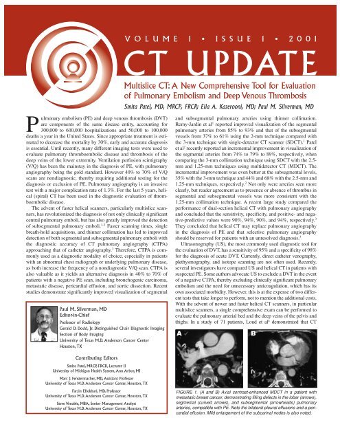
V O L U M E 1 • I S S U E 1 • 2 0 0 1<br />
CT UPDATE<br />
Multislice CT: A New Comprehensive Tool for Evaluation<br />
of Pulmonary Embolism and Deep Venous Thrombosis<br />
Smita Patel, MD, MRCP, FRCR; Ella A. Kazerooni, MD; Paul M. Silverman, MD<br />
and subsegmental pulmonary arteries using thinner collimation.<br />
Remy-Jardin et al 1 reported improved visualization of the segmental<br />
pulmonary arteries from 85% to 93% and that of the subsegmental<br />
vessels from 37% to 61% using the 2-mm technique compared with<br />
the 3-mm technique with single-detector CT scanner (SDCT). 1 Patel<br />
et al 2 recently reported an incremental improvement in visualization of<br />
the segmental arteries from 74% to 79% to 89%, respectively, when<br />
comparing the 3-mm collimation technique using SDCT with the 2.5mm<br />
and 1.25-mm techniques using multidetector CT (MDCT). The<br />
incremental improvement was even better at the subsegmental levels,<br />
35% with the 3-mm technique and 44% and 68% with the 2.5-mm and<br />
1.25-mm techniques, respectively. 2 Not only were arteries seen more<br />
clearly, but reader agreement as to presence or absence of thrombus in<br />
segmental and subsegmental vessels was more consistent with the<br />
1.25-mm collimation technique. A recent large study compared the<br />
performance of dual-section helical CT with pulmonary angiography<br />
and concluded that the sensitivity, specificity, and positive- and negative-predictive<br />
values were 90%, 94%, 90%, and 94%, respectively. 3<br />
They concluded that helical CT may replace pulmonary angiography<br />
in the diagnosis of PE and that selective pulmonary angiography<br />
should be reserved for patients with an unresolved diagnosis. 3<br />
Ultrasonography (US), the most commonly used diagnostic tool for<br />
the evaluation of DVT, has a sensitivity of 95% and a specificity of 98%<br />
for the diagnosis of acute DVT. Currently, direct catheter venography,<br />
plethysmography, and isotope scanning are not often used. Recently,<br />
several investigators have compared US and helical CT in patients with<br />
suspected PE. Some authors advocate US to exclude a DVT in the event<br />
of a negative CTPA, thereby excluding clinically significant pulmonary<br />
embolism and the need for unnecessary anticoagulation, which has its<br />
own associated morbidity. However, this is at the expense of two different<br />
tests that take longer to perform, not to mention the additional costs.<br />
With the advent of newer and faster helical CT scanners, in particular<br />
multislice scanners, a single comprehensive exam can be performed to<br />
evaluate the pulmonary arterial bed and the deep veins of the pelvis and<br />
thighs. In a study of 71 patients, Loud et al 4 demonstrated that CT<br />
A B<br />
FIGURE 1. (A and B) Axial contrast-enhanced MDCT in a patient with<br />
metastatic breast cancer, demonstrating filling defects in the lobar (arrows),<br />
segmental (curved arrows), and subsegmental (arrowheads) pulmonary<br />
arteries, compatible with PE. Note the bilateral pleural effusions and a pericardial<br />
effusion. Mild enlargement of the subcarinal nodes is also noted.
FIGURE 2. Axial contrast-enhanced<br />
MDCT of the pelvis demonstrating<br />
normal optimal enhancement of the<br />
femoral veins (arrows).<br />
venography (CTV) is comparable<br />
with US in the evaluation of DVT<br />
with a sensitivity and specificity of<br />
100%. Garg et al 5 reported obtaining<br />
a good quality CTV examination<br />
in 97% of a total of 70<br />
patients. They demonstrated that<br />
CTV was more efficacious than<br />
US in 36% of cases, equivalent to<br />
US in 37%, and US was better<br />
than CTV in 27%. Duwe et al 6<br />
found an accuracy of 93% for<br />
CTV using US as the reference<br />
test in 74 patients, with a sensitivity of 89%, specificity of 94%, positive-predictive<br />
value of 67%, and negative-predictive value of 98%.<br />
The CTV techniques used by these authors differ, with the scan interval<br />
varying from 5 mm to 20 mm, collimation 5 mm to 10 mm, and the<br />
injection delay from 3 minutes to 3.5 minutes. All of these studies were<br />
performed on a single-detector helical scanner. CTV is particularly useful<br />
in postoperative patients or in obese and immobile patients in whom<br />
sonography can be challenging to perform.<br />
Multi-detector CT Scan Protocol: Part 1–Thorax<br />
For the thorax protocol, 150 cc of iohexol 300 mg/mL<br />
(Omnipaque ® [iohexol]; Amersham Health, Princeton, NJ) is injected<br />
via a 20-gauge angiocatheter in an antecubital vein at a rate of 4<br />
cc/sec. A scan delay of 25 seconds is used. Imaging is performed from<br />
the dome of the diaphragm to the aortic arch during a single breathhold,<br />
with the acquisition time varying between 15 to 19 seconds,<br />
depending on the size of the patient. Images are obtained at 1.25-mm<br />
collimation and reconstructed at 50% of the scan collimation (HS<br />
mode, pitch 6).<br />
Part 2–Leg/pelvic veins<br />
Three minutes following start of the contrast injection, acquisition<br />
of the pelvis and lower extremity veins is obtained from a caudad to<br />
cephalad direction from the level of the tibial plateaus to the iliac<br />
crests. Images are obtained at 7.5-mm collimation and reconstructed at<br />
50% of the scan collimation (HS mode, pitch 6). The entire study is<br />
then evaluated on a GE Advantage Windows Workstation (GE<br />
Medical Systems, Milwaukee, WI). Use of the HS mode in large<br />
patients may account for very noisy images, limiting evaluation of the<br />
pulmonary arteries. In these patients, the HQ mode can be used and<br />
thicker slices obtained. Acquisition of the pulmonary arteries and the<br />
deep veins of the pelvis and thighs is obtained with a single bolus of<br />
intravenous contrast.<br />
Study Interpretation<br />
The window level and width is altered for optimum evaluation of the<br />
pulmonary arteries. A wider window width and a higher level may be<br />
needed to evaluate for subtle emboli. Depending on the concentration<br />
of the contrast in the pulmonary arteries, this may need to be altered at<br />
different levels. Then, the main, lobar, segmental, and subsegmental<br />
arteries are examined systematically from their origins to the 5th or 6th<br />
order branches, using the scrolling mode device. Pulmonary emboli are<br />
diagnosed as partial or complete filling defects, enlargement of the<br />
pulmonary artery with a large occlusive clot, peripheral filling defect,<br />
or nonenhancement of a segment of the pulmonary artery (Figure 1).<br />
Evaluation for chronic pulmonary emboli is also possible, with the<br />
presence of webs or detection of small thick-walled pulmonary arteries<br />
with enhancing walls and filling defects. Systematic review of the<br />
entire pulmonary arterial circulation is performed bilaterally. Note is<br />
also made of additional findings such as pleural or pericardial effusions,<br />
nodules, tumors, nodal enlargement, consolidation, emphysema, and<br />
2<br />
aortic dissection or aneurysm. These aid clinicians in further management<br />
as the clinical presentation of the above diagnoses can be similar.<br />
Then, the veins of the pelvis and thighs are evaluated using the scroll<br />
mode device, after narrowing the window width and level, for optimum<br />
evaluation of the veins.<br />
DVT is diagnosed when there is a central filling defect, enlargement<br />
of the vein (pitfall, venous valves), segmental nonvisualization, wall<br />
enhancement, or perivenous subcutaneous edema (Figures 2 and 3).<br />
Prolonged arterial enhancement may represent extensive bilateral DVT.<br />
However this can also be found when the CT is performed too soon in<br />
the arterial phase or when there is severe bilateral arterial insufficiency.<br />
Observation of other pathology is also possible, such as pelvic tumor<br />
compressing the pelvic veins or a Baker’s cyst. Optimum venous<br />
enhancement is not uniform in all patients with an injection-to-scan<br />
delay of 3 minutes, as this varies according to the patient’s cardiac status<br />
and the state of the pelvic and lower limb arteries. Patients with<br />
altered inflow due to atherosclerotic disease may require a longer delay<br />
time.<br />
Multislice CT allows acquisition of 4 to 8 axial images with each<br />
revolution of the helical scanner, hence accounting for the short scan<br />
acquisition time, which is essential in sick, dyspneic intensive-care<br />
patients who cannot hold their breath. Not only has multislice CT<br />
improved evaluation of the segmental and subsegmental pulmonary<br />
arteries, but it has also allowed evaluation of the lower extremity veins,<br />
the source of PE, with a single injection of intravenous contrast. This<br />
translates into a single imaging modality for a quick, accurate, and efficient<br />
diagnosis of pulmonary thromboembolic disease. Recent studies<br />
have demonstrated
TECHNICAL ISSUES<br />
Abdominal CT Angiography<br />
in the Era of Multidetector CT<br />
Marc J. Fenstermacher, MD; Silvana Faria, MD;<br />
Paul M. Silverman, MD<br />
As new techniques have emerged over the last 15 years, the imaging<br />
evaluation of the vessels of the abdomen has undergone considerable<br />
evolution. Prior to the advent of multidetector CT (MDCT), much<br />
of the advancement has been in magnetic resonance angiography<br />
(MRA). A number of innovative MRA techniques have been utilized,<br />
including 2-dimensional (2D) and 3-dimensional (3D) time-of-flight<br />
(TOF), phase contrast, and, most recently, bolus contrast-enhanced 3D<br />
fast gradient echo techniques, with or without step-wise incremental<br />
couch movement. Currently, the latter approach is widely utilized in<br />
most practices, especially for the evaluation of the arterial and portal<br />
venous systems. The systemic venous system remains a challenge for<br />
accurate, high-contrast MRA, primarily because of the relatively low<br />
concentration of injected gadolinium once it reaches the iliac veins and<br />
inferior vena cava. All of the MRA techniques in the abdomen share a<br />
number of drawbacks, which have fueled the search for better noninvasive<br />
angiographic techniques. With MRA, it is not possible to<br />
demonstrate both the vasculature and the regional soft tissues in the<br />
same imaging sequence; the highest quality arterial MRA will demonstrate<br />
bright vessels and practically nothing else. This problem is particularly<br />
limiting in the oncologic setting, where noninvasive angiography<br />
is used primarily to stage tumors locally and in surgical planning.<br />
Even high-quality abdominal MRA techniques still suffer relatively<br />
low spatial resolution due to the requirement of imaging in the<br />
coronal plane for adequate coverage, with a large field-of-view, yet<br />
avoiding wrap-around aliasing artifacts. Tertiary or smaller branches<br />
are not visualized due to limitations in resolution and slice thickness.<br />
A B<br />
C<br />
D<br />
FIGURE 1. Comparison of magnetic<br />
resonance angiography (MRA) and<br />
computed tomography angiography<br />
(CTA) in a patient with testicular cancer<br />
and a left para-aortic nodal<br />
mass. This preoperative evaluation<br />
was performed prior to retroperitoneal<br />
lymph node dissection. (A)<br />
Coronal MIP projection from 3D<br />
bolus contrast-enhanced MRA in the<br />
arterial phase. (B) Double oblique<br />
coronal CTA reformation showing<br />
two right renal arteries and single left<br />
renal artery. (C and D) Two curved<br />
CTA reformations showing the (C)<br />
upper and (D) lower right renal arteries,<br />
and the left para-aortic nodal<br />
mass abutting the undersurface of<br />
the left renal artery.<br />
3<br />
Any patient motion during the 20- to 30-second breath-hold acquisition<br />
degrades the MRA images severely.<br />
With single-detector helical CT (SDCT), it first became possible to<br />
reconstruct the 3D helix retrospectively with the acquired slice thickness<br />
but with much smaller intervals, such as 50% of the collimation.<br />
This enabled the creation of high-quality multiplanar volume reformations<br />
and other angiographic images using a workstation, and we began<br />
using this technique in selected cases for visualizing the arterial and<br />
venous vasculature adjacent to tumors in the abdomen and pelvis. The<br />
limiting factors with SDCT have been the interrelated issues of slice<br />
thickness and coverage. All bolus-contrast-enhanced angiographic<br />
techniques depend on matching the timing of image acquisition to the<br />
transit of contrast through the vessels of interest. Since all acquisitions<br />
are in the axial plane, there is limited coverage with the 2- to 3-mm collimation<br />
and pitch of 2 usually used with SDCT. 1<br />
The advent of MDCT has allowed the acquisition of larger areas of<br />
coverage with thinner slice collimation and much faster imaging<br />
times per series. We routinely image the entire abdominal aorta and<br />
proximal common iliac arteries in
transit of the bolus of contrast through the aorta. An alternative to<br />
SmartPrep, which does incur a nominal though mandatory delay<br />
between contrast arrival in the abdomen and the initiation of scanning,<br />
is to perform a test injection in order to estimate the contrast travel<br />
time to the abdominal aorta. This step is time consuming and not used<br />
routinely in our department at this time.<br />
Data is transferred to a workstation, currently a Vital Images<br />
Vitrea2 (Vital Images, Inc., Minneapolis, MN) or an Advantage<br />
Windows workstation (GE Medical Systems), for creating and filming<br />
curved and multiple oblique reformations. Although this is a timeconsuming<br />
step for the radiologist, it is rewarding for the clinician.<br />
While a technologist with special interest and training in image processing<br />
can perform this step in many cases, optimizing reformatted<br />
imaging planes often requires a knowledge of anatomy and pathology<br />
that may be beyond their reach (Figure 2). At the current stage of<br />
development of these techniques, interested cross-sectional radiologists<br />
should be encouraged to perform the reformations and recon-<br />
PRACTICAL ISSUES<br />
Imaging Considerations in Pediatric<br />
Multislice CT: A Radiologist’s Perspective<br />
Farzin Eftekhari, MD and Paul M. Silverman, MD<br />
Multislice CT, occasionally referred to as multidetector or multidetector<br />
row CT, has made a significant impact on the imaging of<br />
pediatric patients by reducing the total study time in uncooperative or<br />
very ill patients. In many instances, this has virtually eliminated<br />
breathing or motion artifacts, and, even more importantly, the need for<br />
conscious sedation or general anesthesia. 1<br />
In those patients who require conscious sedation or general anesthesia,<br />
shorter examination time has minimized the sedation period,<br />
and, thus, the potential risks of sedation or anesthesia. This has<br />
increased patient throughput and the efficiency of the sedation team,<br />
enabling them to schedule additional pediatric CT, magnetic resonance<br />
(MR), or interventional radiology procedures.<br />
Table 1. Technical Considerations in Pediatric Multislice CT<br />
Approximate Approximate<br />
DFOV mA kV Age (y) Weight (kg)<br />
processes and to cover the smaller anatomy.<br />
It is also important to realize that the source-to-detector distance is<br />
decreased in the LightSpeed multislice scanner (94.9 cm) relative to<br />
the GE single-slice design (109.9 cm). This detector alone accounts for<br />
a 34% increase in radiation dose for the multislice system compared<br />
with the single-slice configuration using identical scan parameters.<br />
For pediatric abdominal/pelvic CT scanning, we use a slice thickness<br />
of 5 mm, high speed (HS), and a table speed of 22.5 mm/rotation.<br />
For pediatric chest CT, we use a slice thickness of 5 mm, HS,<br />
and 22.5 mm/rotation table speed.<br />
Pediatric patients receive iodinated contrast material when it is<br />
clinically indicated. To limit reactions or nausea and vomiting, we use<br />
low-osmolality contrast material (LOCM) at a dose of 1.5mL/kg<br />
body weight for the chest, and 2.0 mL/kg for abdominal and pelvic<br />
CT. We prefer to deliver all of the contrast before scanning and allow<br />
a 25-second delay for chest, and chest/abdomen/pelvis imaging<br />
(allowing a 12-second pause between chest and abdomen), and 50<br />
seconds for abdomen/pelvis imaging. Alternatively, when available,<br />
we use a computer automated scanning technology, such as<br />
SmartPrep, to ensure that scanning occurs during peak liver enhancement.<br />
This allows for consistency and optimal imaging to detect focal<br />
liver lesions.<br />
Injection rates for contrast material range from 0.3 to 3.0mL/sec,<br />
depending on the size of the IV access line (22- to 18-gauge angiocatheters,<br />
depending on the age of the child).<br />
Our pediatric technique (Table 1) is based on the child’s body size<br />
(depth of field of view), age, and weight. For children older than 6<br />
years of age, we use a slice thickness of 7.5 mm and 15 mm/rotation<br />
table speed. We use this table to select the appropriate mA setting for<br />
each case. The mA ranges from 140 to 230 with a kV of 120. This<br />
MEDICAL ECONOMICS<br />
Application of Six Sigma to Healthcare:<br />
Improving CT Efficiency<br />
Steve Venable, MBA and Paul M. Silverman, MD<br />
Six Sigma is a performance improvement methodology used to<br />
achieve significant change and improvement. The term Six Sigma<br />
refers to six standard deviations from the mean on a normal distribution<br />
model. This level of performance would seem impossible to attain at<br />
first, but the methodology leads to extraordinary improvement on<br />
processes and final results. Many large corporations, including Allied<br />
Signal, Honda, and GE, 1 have adopted Six Sigma to improve their<br />
financial performance and maintain a competitive advantage.<br />
Six Sigma represents a cultural shift to continuous, data-driven<br />
performance improvement. Though Six Sigma was originally used to<br />
decrease defects in manufacturing industries, it has moved to service<br />
industries, and is readily adapted to healthcare. The data-driven<br />
aspect of Six Sigma meshes well in the current hospital setting where<br />
dramatic efforts are being made to strive for marginal efficiency in a<br />
competitive environment. Physicians require a business methodology<br />
based on verifiable data and analysis. This focuses all groups on solutions<br />
to problems and quantitative measures of success.<br />
At the University of Texas M.D. Anderson Cancer Center<br />
(MDACC), we have used Six Sigma to provide a dramatic improvement<br />
in efficiency. In computed tomography (CT), a common reason<br />
given for patient delays was a lack of laboratory tests prior to the CT<br />
scan. More than 95% of our patients received IV contrast and labs<br />
were required prior to administering contrast. However, data analysis<br />
5<br />
table is designed for current GE multislice scanners in which the scan<br />
time is 0.8 seconds. However, since newer scanners offer shorter scan<br />
times, such as 0.5 seconds, we intend to update this table to adjust for<br />
mA values accordingly.<br />
Conclusion<br />
We feel that the use of multislice CT scanners has had a significant<br />
impact on imaging in pediatric patient population in terms of need for<br />
sedation and throughput, but we need to continue to look for ways to<br />
decrease the radiation dose to even lower levels.<br />
Acknowledgments<br />
The authors would like to thank Dianna D. Cody, PhD, and Donna<br />
M. Moxley, MS, from Diagnostic Physics for their valuable comments.<br />
REFERENCES<br />
1. Pappas JN, Donnelly LF, Frush DP. Reduced frequency of sedation of young children<br />
with multisection helical CT. <strong>Radiology</strong>. 2000;215:897-899.<br />
2. McCollough CH, Zink FE. Performance evaluation of a multislice CT system. Med<br />
Phys. 1999;26:2223-2230.<br />
3. Cody DD, Moxley DM, Eftekhari F, Silverman PM. Pediatric dose considerations<br />
in multislice helical CT. Helical CT Today. 2001;6(3):3-4.<br />
4. Motley DM, Hazle JD, Shepard JS, Zhou XJ. Dosimetry of a new multislice helical<br />
CT scanner: Calculated and measured patient doses [abstract]. <strong>Radiology</strong>.<br />
1999;213(P):284.<br />
5. Donnelly LF, Frush DP, Nelson RC. Multislice helical CT to facilitate combined CT<br />
of the neck, chest, abdomen, and pelvis in children. AJR Am J Roentgenol.<br />
2000;174:1620-1622.<br />
6. Brenner DJ, Elliston CD, Hall EJ, Berdon WE. Estimated risks of radiationinduced<br />
fatal cancer from pediatric CT. AJR Am J Roentgenol. 2001;176:289-296.<br />
7. Paterson A, Frush DP, Donnelly LF. Helical CT of the body: Are settings adjusted<br />
for pediatric patients? AJR Am J Roentgenol. 2001;176:297-301.<br />
found that
measured. We discovered that many long-held perceptions were inaccurate.<br />
Perception: “Many” patients reported without the requisite<br />
labs; Reality: 3 minutes, even for a combined chest, abdomen,<br />
and pelvis exam. We spent more time getting the patient onto and off<br />
the table, and the setup and cleaning of the room, 20 minutes total.<br />
The team concentrated on optimizing the patient-related activities and<br />
removed the excess idle time of the CT scanners.<br />
The Interpretive Process<br />
The teams also analyzed the activities in the reading rooms. The<br />
goal was to eliminate all nonvalue activities performed by the radiologists<br />
and shift those activities to others, allowing the radiologists to<br />
concentrate on film interpretation and reports. We began to print out<br />
the information needed by the radiologists for protocoling the patients<br />
putting pertinent information at their fingertips. We also began to hang<br />
the motorized viewers continuously throughout the day, relieving the<br />
radiologist of this tedious task. The results were a dramatic increase in<br />
throughput in the reading rooms and the radiologists were able to stay<br />
current with the growing patient activity.<br />
During the characterization process, several processes were determined<br />
not to need the full Six Sigma treatment, instead, a small group<br />
could work out a solution. An example was the 50% turnover in CT<br />
technologists. A working group interviewed the technologists and<br />
analyzed external market issues. It was determined that the techs were<br />
undercompensated compared with the local market and there were<br />
inconsistencies in the work environment. Work volumes between<br />
shifts and lack of communication between technologists on each shift<br />
were problematic. As soon as changes were implemented to correct<br />
these issues, turnover was reduced to 0% the following year.<br />
Conclusion<br />
When this Six Sigma project was initiated, we failed to realize its<br />
size and scope. We also underestimated the manpower resources<br />
required. Six Sigma touches the entire operation and the necessary<br />
resources must be dedicated to the project. Cultural changes and the<br />
fear of change must be overcome. Six Sigma is data rigorous and provides<br />
a framework for disciplined analysis and correction of processes<br />
not meeting the specification. The payoffs are tremendous for the<br />
corporate culture, for patient satisfaction, and in increased throughput<br />
(shortened patient wait times, faster exam times, decreased interpretation<br />
time resulting), with financial benefits in the increased capacity<br />
on the same resources. For the radiologist, we removed the nonvalue-added<br />
activities in the reading rooms and developed closer working<br />
relationships between the physician staff and technical and nursing<br />
staff. Weaknesses in our information systems were identified and<br />
detailed changes to the software were initiated. Our film printing system<br />
was woefully inadequate and a new system was designed, purchased,<br />
and installed to improve the film quality control function, add<br />
more capacity, and remove the film printing bottleneck. The financial<br />
returns for this project are projected at $15.8 million over 5 years. For<br />
every dollar invested in this project, MDACC expects $16 in return.<br />
The final lesson is that Six Sigma is never-ending; we monitor and<br />
improve our processes continuously. As new areas come into focus, we<br />
apply the tools and continue to reap the benefits of the program.<br />
REFERENCES<br />
1. Harry MJ, Schroeder R. Six Sigma: The Breakthrough Management Strategy Revolutionizing<br />
the World’s Top Corporations. pp. vii-viii: 112. New York: Random House,<br />
2000.<br />
2. Harry MJ, Lawson JR. Six Sigma Producibility Analysis and Process<br />
Characterization. pp. 4.2-4.4. Boston: Addison-Wesley Publishing Co., 1992.<br />
FUTURE MEETINGS AND CONVENTIONS<br />
Meeting date Meeting name Location Contact<br />
November 1-8, 2001 American Society for Therapeutic <strong>Radiology</strong> and Oncology San Francisco, CA 703-502-1550<br />
November 25-29, 2001 Radiological Society of North America Chicago, IL 630-571-2670<br />
CT Update is published by Anderson Publishing, Ltd., 1301 West Park Ave., Ocean, NJ 07712 • (732) 695-0600.<br />
O. Oliver Anderson, Publisher; Elizabeth A. McDonald, Managing Editor; Felice Ponger, Art Director.<br />
Sponsored by an educational grant from Amersham Health. The views and opinions expressed in this publication are those of the authors and do not necessarily<br />
reflectthose of the publisher or sponsor. Full and complete prescribing information should be reviewed regarding any product mentioned prior to use.<br />
6