Leprosy Training Module for Medical Officers
Leprosy Training Module for Medical Officers
Leprosy Training Module for Medical Officers

Create successful ePaper yourself
Turn your PDF publications into a flip-book with our unique Google optimized e-Paper software.
N<br />
The Causative Organism – M. leprae<br />
<strong>Leprosy</strong> is caused by Mycobacterium leprae, which was discovered<br />
in 1873 by Hansen at Bergen in Norway. These Mycobacterium leprae<br />
are pleomorphic, straight or slightly curved, rod – shaped gram positive<br />
micro - organisms. They may appear like solid rods, fragmented or<br />
granular. They are acid fast bacilli ( AFB ), because they are stained red<br />
by a dye called carbol fuschin , and this red colour can not be removed<br />
either by acid or alcohol. M. leprae is less acid fast than M. tuberculosis.<br />
The presence of the bacilli can be demonstrated by taking skin<br />
smears, and after staining with Zeihl Neilson stain , these bacilli can be seen<br />
under microscope. They are seen lying singly, in clumps or in compact<br />
masses known as globi. The living <strong>Leprosy</strong> bacilli appear as solid staining,<br />
i.e., bright pink rods with rounded ends and uni<strong>for</strong>mly stained through<br />
their entire length. Although the discovery of <strong>Leprosy</strong> bacilli reported as early<br />
as 1873, they could not yet be grown in artificial culture media. The generation<br />
time of M. leprae in the mouse footpad is 12 – 14 days ( longest of any known<br />
bacterium ). In comparison with this, the generation time of M. tuberculosis is<br />
only 20 hours.<br />
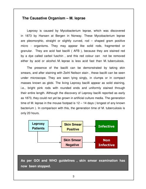
<strong>Leprosy</strong><br />
Patients<br />
Skin Smear<br />
Positive<br />
Infective<br />
Skin Smear<br />
Negative<br />
o n<br />
Infective<br />
As per GOI and WHO guidelines , skin smear examination has<br />
now been stopped.<br />
3