Whipple's disease and Tropheryma whipplei
Whipple's disease and Tropheryma whipplei
Whipple's disease and Tropheryma whipplei
You also want an ePaper? Increase the reach of your titles
YUMPU automatically turns print PDFs into web optimized ePapers that Google loves.
SUMMARY : Whipple’s <strong>disease</strong> <strong>and</strong> <strong>Tropheryma</strong><br />
<strong>whipplei</strong><br />
-Introduction.<br />
-What about the micro-organism?<br />
-Habitat, source <strong>and</strong> transmission of T. <strong>whipplei</strong>.<br />
-Risk factors?<br />
-Clinical signs <strong>and</strong> symptoms of Whipple’s <strong>disease</strong>.<br />
-Samples for the diagnosis of Whipple’s <strong>disease</strong>.<br />
-Non specific diagnosis.<br />
-Specific diagnosis.<br />
-Interpretation of results.<br />
-Treatment of the <strong>disease</strong>?<br />
-Counter-indications of treatment?<br />
-Secondary effects of treatment?<br />
-Evolution <strong>and</strong> prognosis of the <strong>disease</strong>?<br />
-Follow-up of the treated patients?<br />
-Follow-up of the patients after the end of the treatment?<br />
-List of the publications of IFR 48 about Whipple’s <strong>disease</strong>.<br />
-Interlocutors in IFR 48.<br />
-Consultation at IFR 48.<br />
-What kind of samples to send <strong>and</strong> how ?<br />
-Sheet of clinical information.<br />
-Information for the patients.
Introduction<br />
Whipple’s <strong>disease</strong> is an infectious <strong>disease</strong> caused by a<br />
bacterium, <strong>Tropheryma</strong> <strong>whipplei</strong>. The <strong>disease</strong> is rare (annual<br />
incidence : one per million habitant) but its prevalence is not<br />
really known.<br />
The <strong>disease</strong> has been described for the first time in 1907<br />
by an American physician, George Whipple.<br />
The clinical manifestations are various <strong>and</strong> are not really<br />
specific leading to a delay for the diagnosis.<br />
The spontaneous evolution of the <strong>disease</strong> is long,<br />
marked by remission <strong>and</strong> relapse episodes, that could be<br />
evolved until the death in absence of specific treatment.<br />
T. <strong>whipplei</strong> has been considered for a long time as<br />
uncultivable. The first isolation <strong>and</strong> establishment of a strain of<br />
T. <strong>whipplei</strong> has been performed in our laboratory in 1999 from a<br />
cardiac valve of a patient with an endocarditis due to T.<br />
<strong>whipplei</strong>.<br />
This step has been determinant for new perspectives of<br />
diagnosis <strong>and</strong> treatment of the <strong>disease</strong>.<br />
References:<br />
-Whipple’s <strong>disease</strong>. Marth T, <strong>and</strong> D Raoult. Lancet 2003: 18, 239-246.<br />
-Whipple’s <strong>disease</strong> <strong>and</strong> “<strong>Tropheryma</strong> whippelii”. Dutly F, <strong>and</strong> M Altwegg.<br />
Clin Microbiol Rev 2001: 14, 561-583.
What about the micro-organism?<br />
The cell-culture of the bacterium on human fibroblasts<br />
has allowed its characterization.<br />
Phylogenetical analysis has classified the bacterium<br />
among the Gram positive bacterium at high GC%, near 2<br />
species pathogens for human, Actinomyces pyogenes <strong>and</strong><br />
Rothia dentocariosa <strong>and</strong> other bacteria from the environment.<br />
The culture of this bacterium has also allowed the full<br />
sequencing of the genome that has been performed in parallel<br />
on 2 distinct strains.<br />
The genome analysis has shown the presence of repeated<br />
sequences which could allow genomic recombination of T.<br />
<strong>whipplei</strong> <strong>and</strong> could modify the expression of membrane<br />
protein.<br />
A deficiency in the synthesis of amino-acids has been<br />
also observed during the genome analysis. To compensate this<br />
deficit, we have developed a medium culture supplemented with<br />
all the amino-acids, that have allowed the growth of T. <strong>whipplei</strong><br />
in axenic conditions.<br />
References :<br />
-<strong>Tropheryma</strong> <strong>whipplei</strong> Twist: a human pathogenic Actinobacteria with a<br />
reduced genome. Raoult et al. Genome Res 2003: 13:1800-9.<br />
-Genome-based design of a cell-free culture medium for <strong>Tropheryma</strong><br />
<strong>whipplei</strong>. Renesto et al. Lancet 2003: 362:447-9.
Phylogeny of <strong>Tropheryma</strong> <strong>whipplei</strong> using the 16S<br />
rRNA sequence<br />
Eubacterium lentum<br />
Gardnerella vaginalis<br />
Mobiluncus spp.<br />
<strong>Tropheryma</strong> <strong>whipplei</strong><br />
Rothia dentocariosa<br />
Actinomyces spp.<br />
Propionibacterium spp.<br />
Corynebacterium spp.<br />
Nocardia spp.<br />
Rhodococcus spp.<br />
Gordona spp.<br />
Mycobacterium spp.
Habitat, source, <strong>and</strong> transmission of T. <strong>whipplei</strong><br />
The source of T. <strong>whipplei</strong> <strong>and</strong> its transmission are<br />
presently unknown. The bacterium seems to be present in the<br />
environment. A study based on PCR has shown the presence<br />
of T. <strong>whipplei</strong> DNA in sewage water.<br />
One hypothesis could be that the transmission was linked<br />
to an oro-fecal route. Several points support this hypothesis :<br />
the presence of the bacterium in sewage water, the possible<br />
transmission of the bacterium in association with Giardia<br />
intestinalis or by digestive fibroscopy.<br />
The Whipple’s <strong>disease</strong> is ubiquitous. It seems however,<br />
more frequent in some geographical area, mainly, for France<br />
in Rhône-Alpes area, but also in Switzerl<strong>and</strong> <strong>and</strong> Germany.<br />
References:<br />
-Whipple’s <strong>disease</strong>. Marth T, <strong>and</strong> D Raoult. Lancet 2003: 18,<br />
239-46.<br />
-Can Whipple’s <strong>disease</strong> be transmitted by gastroscopes? La<br />
Scola et al. Infect Control Hosp Epidemiol 2003: 24, 191-4.<br />
-Whipple’s <strong>disease</strong> associated with giardiasis. Fenollar et al. J<br />
Infect Dis 2003: 188, 828-34.
Risk factors?<br />
Certain studies underlined the possible existence of<br />
specific factors to develop Whipple’s <strong>disease</strong>. Indeed, men of<br />
50 years olds are the more frequently involved. The <strong>disease</strong><br />
seems also to be more frequent among farmers.<br />
Different immunological abnormalities have been<br />
reported during Whipple’s <strong>disease</strong> in treated patients but the<br />
data have been contradictory.<br />
Recently, a diminution of production of Interleukine-12<br />
<strong>and</strong> IFN-γ by periphereal monocytes has been shown for<br />
patients with Whipple’s <strong>disease</strong> in comparison to control people,<br />
not only in acute phase but also in remission. It is necessary<br />
now to confirm these data.<br />
References :<br />
-Defects of monocyte interleukin-12 production <strong>and</strong> humoral<br />
immunity in Whipple’s <strong>disease</strong>. Gastroenterology 1997 : 113 :<br />
442-448.<br />
-Whipple’s <strong>disease</strong>. Marth T, <strong>and</strong> D Raoult. Lancet 2003: 18,<br />
239-246.
Clinical manifestations of Whipple’s <strong>disease</strong><br />
For a long time, the Whipple’s <strong>disease</strong> has been<br />
considered as a digestive <strong>disease</strong>. In reality, the clinical<br />
manifestations are various <strong>and</strong> non specific. Approximately<br />
15% of the patients do not present digestive signs. The <strong>disease</strong><br />
begins most often, by arthralgia. Presently, we could distinguish<br />
3 clinical presentations :<br />
-The classic Whipple’s <strong>disease</strong> is multivisceral. Patient<br />
present digestive symptoms (mainly diarrhea), arthralgia <strong>and</strong>/ or<br />
adenopathies eventually associated to brain or cardiac<br />
involvement. The digestive biopsy is PAS-positive.<br />
-The blood-culture negative endocarditis, without<br />
digestive involvement, for which the diagnosis is particularly<br />
difficult, as in most documented cases, it has been performed<br />
on cardiac valves analysis.<br />
-The isolate neurological infection, without digestive<br />
involvement, for which the diagnosis is particularly difficult. A lot<br />
of neurological signs could be observed such as cognitive<br />
impairment, ocular paralysis, abnormal ocular movements,<br />
myoclonus, <strong>and</strong>/or hypothalamic involvement.<br />
References:<br />
-Whipple’s <strong>disease</strong>. Marth T, <strong>and</strong> D Raoult. Lancet 2003: 18, 239-246.<br />
-Whipple’s <strong>disease</strong>. Fenollar F, <strong>and</strong> D Raoult. Clin Diagn Lab Immunol 2001 : 8, 1-7.
Samples for the diagnosis of Whipple’s <strong>disease</strong><br />
In case of suspicion of Whipple’s <strong>disease</strong>, saliva, stools,<br />
blood <strong>and</strong> cerebrospinal fluid (CSF) could be easily sampled<br />
for PCR <strong>and</strong> culture. Small-bowel biopsies must be performed<br />
even of case of lack of digestive signs. In function of the<br />
symptoms, other biopsies could be performed for PAS-staining<br />
immunohistochemistry <strong>and</strong> PCR. Finally, if a final diagnosis of<br />
Whipple’s <strong>disease</strong> is performed, a CSF should be<br />
systematically sampled for a PCR because an asymptomatic<br />
neurological involvement has been described.<br />
-Specimen depending of the clinical manifestations :<br />
Classic WD Endocarditis Neuro-Whipple<br />
Saliva Systematic (S) S S<br />
Stools S S S<br />
Blood S S S<br />
CSF<br />
To perform in case To perform in case S<br />
Small-bowel<br />
of definite diagnosis of definite<br />
of WD<br />
diagnosis of WD<br />
S S S<br />
biopsy<br />
Cardiac valve Not necessary (NN) 2 2<br />
Adenopathy, Second intention 2 2<br />
synovial biopsy exam (2)<br />
Brain biopsy NN NN Third intention<br />
References:<br />
-Whipple’s <strong>disease</strong>. Marth T, <strong>and</strong> D Raoult. Lancet 2003: 18, 239-46.<br />
exam (3)<br />
-Molecular techniques in Whipple’s <strong>disease</strong>. Expert Rev Mol Diagn 2001: 1, 299-309.<br />
Non specific diagnosis
The hemogram could show an anemia. An eosinophilia<br />
could be also observed as ESR or CRP increasing. Biological<br />
signs of digestive malabsorption could be present.<br />
The analysis using Periodic Acid Schiff (=PAS)<br />
staining could be performed on various biopsies (classically<br />
on small-bowel biopsies) <strong>and</strong> also in body fluid to look for the<br />
presence of macrophages with positive PAS material.<br />
Positive PAS-staining from a duodenal biopsy. (x 100).<br />
References:<br />
-Whipple’s <strong>disease</strong>. Marth T, <strong>and</strong> D Raoult. Lancet 2003: 18, 239-246.<br />
-Whipple’s <strong>disease</strong>: immunospecific <strong>and</strong> quantitative immunohistochemical study of<br />
intestinal biopsy specimen. Lepidi et al. Human Pathol 2003: 34: 589-96..
Specific diagnosisThe specific diagnosis includes :<br />
Immunohistochemistry<br />
Genomic amplification by PCR<br />
Culture<br />
The immunohistochemistry with polyclonal rabbit<br />
antibodies specifically directed against the bacterium could be<br />
performed on different samples (small-bowel, cardiac valve,<br />
brain, adenopathy), as body fluids (articular fluid, aqueous<br />
humor) <strong>and</strong> blood. This technique should be retrospectively<br />
performed on formalin-fixed samples.<br />
Positive immunohistochemistry of a duodenal biopsy. (x 100).<br />
-References :<br />
-Diagnosis of Whipple <strong>disease</strong> by immunohistochemical analysis: a sensitive <strong>and</strong><br />
specific method for the detection of T. <strong>whipplei</strong> in paraffin-embedded tissue. Baisden<br />
et al. Am J Clin Pathol 2002: 118, 742-8.<br />
-T. <strong>whipplei</strong> circulating in blood monocytes. Raoult et al. N Engl J Med 2001: 345,<br />
548.<br />
-Immunohistological detection of T. <strong>whipplei</strong> in lymph nodes. Lepidi et al. Am J Med<br />
2002:118, 742-8.<br />
-Immunodetection of T. <strong>whipplei</strong> in intestinal tissues from Dr Whipple’s 1907 patient.<br />
Dumler et al. N Engl J Med 2003: 348:1411-2.
Genomic amplification by PCRPCR could be performed<br />
on various biopsies, body fluids, blood, stools or saliva<br />
using primers targeting various genes of the bacterium. The<br />
analysis of complete genome of the bacterium has allowed a<br />
rationalisation for the choice of primers. A real-time PCR<br />
targeting repeated sequences of T. <strong>whipplei</strong> has allowed an<br />
increasing of sensitivity without altering the specificity of the<br />
technique.<br />
T. <strong>whipplei</strong> DNA has been detected in saliva, small-bowel<br />
biopsies, gastric fluid <strong>and</strong> stools of healthy people. However,<br />
these data should be interpreted cautiously as they are based<br />
on « home-made » PCR <strong>and</strong> have never been confirmed by<br />
other teams. The exact prevalence of asymptomatic carriers<br />
of T. <strong>whipplei</strong> need still to be determined. The problem with<br />
PCR is the risk of contamination. To conclude to the positivity of<br />
a sample, it is necessary to obtain 2 positive PCR targeting 2<br />
different DNA sequences.<br />
Culture<br />
The culture from biopsies (duodenum, cardiac valve…),<br />
body fluids (CSF) or blood could be performed using cell<br />
culture technique or axenic medium supplemented with amino<br />
acids.<br />
References:<br />
-Use of genome selected repeated sequences increases the sensitivity of PCR detection of T.<br />
<strong>whipplei</strong>. Fenollar et al. J Clin Microbiol 2004: 42, 401-3.<br />
-Culture of T. <strong>whipplei</strong> from human samples: a 3-year experience (1999 to 2002). J Clin Microbiol<br />
2003: 41, 3816-22.
Primers targeting specifically <strong>Tropheryma</strong> <strong>whipplei</strong> published in the<br />
litterature.<br />
Primers<br />
Sens/<br />
Sequences<br />
Sens/Antisens<br />
Targeted<br />
gene<br />
Antisens<br />
tws3,f/<br />
tw1857r1<br />
tw1662f/<br />
tw1857r1<br />
tws1,f/<br />
tws2,r<br />
tws3,f/<br />
tw1857r1<br />
tws3,f/<br />
tws4,r<br />
TW-23InsF/<br />
TW-23InsR2<br />
whipp-frw1/<br />
whipp-rev<br />
whipp-frw2/<br />
whipp-rev<br />
TwrpoB.F/<br />
TwrpoB.R<br />
53.3F/<br />
5’CCGGTGACTTAACCTTTTTGGAGA3’/<br />
5’TCCCGAGCCTTTCCGAG A3’<br />
5’ACTATTGGGTTTTGAGAGGC3’/<br />
5’TCCCGAGCCTTATCCGAG A3’<br />
5’ATCGCAAGGTGGAGCGAATCT3’/<br />
5’CGCATTCTGGCGCCCCAC3’<br />
5’CCGGTGACTTAACCTTTTTGGAGA3’/<br />
5’TCCCGAGCCTTATCCGAGA3’<br />
5’CCGGTGACTTAACCTTTTTGGGA3’/<br />
5’CTCCCGTGAGCTTGTGCCCAAAC3’<br />
5’GGTTGATATTCCCGTACCGGCAAAG3’/<br />
5’GCATAGGATCACCAATTTCGCGCC3’<br />
5’TGACGGGACCACAACATCTG3’/<br />
5’ACATCTTCAGCAATGATAAGAAGTT3’<br />
5’CGCGAAAGAGGTTGAGACTG3’<br />
5’ACATCTTCAGCAATGATAAGAAGTT3’<br />
5’AAAAAGGCCGCACGCGAGTT’/<br />
5’AAAGAGGCTCCAACGCCACG3’<br />
5’AGAGAGATGGGGTGCAGGAC3’/<br />
ITS<br />
ITS<br />
ITS<br />
ITS<br />
ITS<br />
23S rDNA<br />
hsp65<br />
hsp65<br />
rpoB<br />
Repeat<br />
53.3R 5’AGCCTTTGCCAGACAGACAC3’<br />
References:<br />
-Molecular genetic methods for the diagnosis of fastidious microorganisms. Fenollar<br />
F, <strong>and</strong> D Raoult. APMIS 2004; 112:785-807.
Interpretation of results ? When a patient presents a<br />
« classic » <strong>disease</strong> with digestive signs, adenopathies <strong>and</strong>/or<br />
arthralgia, the diagnosis is easy. In case of <strong>disease</strong> without<br />
digestive involvement (endocarditis, neurologic<br />
involvement…), the diagnostic is more difficult. One of the<br />
problems is to interpret an isolated positive PCR on blood or<br />
CSF. These results should always be confirmed. In these cases,<br />
the presence of PCR + on saliva or stools could be useful to<br />
confirm the diagnosis.<br />
-Strategy of interpretation of the results in function of samples <strong>and</strong><br />
techniques :<br />
Specimen PAS IHC PCR Diagnostic +<br />
Small bowel + - or NE - or NE NO<br />
Adenopathy + NE + YES<br />
Cardiac Valve - or NE + + YES<br />
Brain - or NE - or NE + YES (?)<br />
Other biopsies - or NE + - or NE YES (?)<br />
CSF + - or NE - or NE NO<br />
Blood + - or NE + YES<br />
- or NE + +<br />
YES - or NE - or NE<br />
+ ?*<br />
Saliva NE NE + Predictive value<br />
depending of the laboratory<br />
Stools<br />
* Necessity to confirm on a second specimen, eventually targeting a new DNA<br />
sequence ; NP: Not performed, -: negative; +: positive
-References: Molecular genetic methods for the diagnosis of fastidious<br />
microorganisms. Fenollar F, <strong>and</strong> D Raoult. APMIS 2004; 112:785-807.
Treatment of Whipple’s <strong>disease</strong>? -Whipple’s <strong>disease</strong><br />
without neurologic involvement (Negative PCR on CSF <strong>and</strong><br />
absence of neurologic symptoms)<br />
Doxycycline, 100 mg, twice per day.<br />
In association with<br />
Hydroxychloroquine, 200 mg, three times per day.<br />
This association is administered for a length of 18 months at<br />
least.<br />
-Whipple’s <strong>disease</strong> with neurologic involvement<br />
Doxycycline, 100 mg, twice per day.<br />
In association with<br />
Hydroxychloroquine, 200 mg, three times per day In<br />
association with<br />
Sulfamethoxazole <strong>and</strong> Trimethoprim, 1,600 mg <strong>and</strong> 320 mg,<br />
three times per day OR Sulfadiazine, 1,500 mg, four times per<br />
day<br />
This association is administered for a length of 18 months at<br />
least.<br />
Moreover : Add folic acid: 10 mg per day for the patients > 65<br />
years <strong>and</strong> patients with deprive.<br />
References:<br />
-Antibiotic susceptibility of <strong>Tropheryma</strong> <strong>whipplei</strong> in MRC5 cells. Boulos et al.<br />
Antimicrob Agents Chemoter 2004: 48, 747-752.<br />
-Molecular evaluation of antibiotic susceptibility against <strong>Tropheryma</strong> <strong>whipplei</strong> in<br />
axenic medium. Boulos et al. J Antimicrob Chem 2005: 2, 178-181.
Counter-indications of treatment ?<br />
-Doxycycline<br />
Allergy to tetracyclines.<br />
Child of less than 8 years.<br />
Pregnant woman or woman who nurses.<br />
-Hydroxychloroquine<br />
Allergy to hydroxychloroquine.<br />
Retinopathy.<br />
-Cotrimoxazole <strong>and</strong> Sulfadiazine<br />
Allergy to sulfamides.<br />
Deficit in Glucose-6-phosphate Dehydrogenase.
Secondary effects of treatment?<br />
-Doxycycline<br />
Photosensitization (It is necessary to protect the face <strong>and</strong> the<br />
h<strong>and</strong>s of sun, mainly the summer).<br />
Digestive troubles, allergy, hematological troubles, cutaneous<br />
pigmentation.<br />
-Hydroxychloroquine<br />
Photosensitization, acouphene, giddiness, digestive troubles,<br />
cutaneous reaction, headache, hematological troubles,<br />
retinopathy, psychoses.<br />
-Cotrimoxazole <strong>and</strong> Sulfadiazine<br />
Allergy, hematological troubles, digestive troubles, hepatitis,<br />
pseudo-membranous colitis, alteration of kidney function,<br />
urinary lithiasis, aseptic meningitis.
Evolution <strong>and</strong> prognosis of the <strong>disease</strong>?<br />
In lack of adapted antibiotic treatment, the evolution of<br />
Whipple’s <strong>disease</strong> is fatal.<br />
For the patients treated adequately, the diarrhea<br />
disappeared quickly, in less of one week. The arthralgia<br />
regress between 2 to 3 weeks. For neurological signs, their<br />
regression is generally observed, more than 3 weeks after the<br />
beginning of the adapted antibiotic treatment. Moreover,<br />
neurological sequellaes are frequently observed.<br />
After the end of the antibiotic treatment, relapses are<br />
observed, mainly, in the brain. Their frequency has been<br />
diversely estimated between 2 <strong>and</strong> 35%.<br />
After the end of the antibiotic treatment, it is better to<br />
follow the patients during his entire life.<br />
References:<br />
-Whipple’s <strong>disease</strong>. Marth T, <strong>and</strong> D Raoult. Lancet 2003: 18, 239-246.
Follow-up of the treated patients?<br />
-Monthly :<br />
Hemogram, transaminases, gammaglutamyltranspeptidase <strong>and</strong><br />
creatinine blood levels.<br />
Immunohistochemistry of monocytes, saliva, blood <strong>and</strong> stools<br />
for PCR.<br />
Plasmatic levels of prescribed antibiotics to check for the<br />
presence of therapeutic levels:<br />
Hydroxychloroquine : 1 µg/ml<br />
Doxycycline : 4 µg/ml<br />
Sulfamethoxazole or sulfadiazine : 100 µg/ml<br />
Necessity to perform, 2 months after the beginning of<br />
antibiotic therapy for patients with neurological<br />
involvement :<br />
Lumbar puncture for PCR <strong>and</strong> antibiotics levels<br />
(doxycycline, sulfamethoxazole or sulfadiazine) in CSF.<br />
-Semi-annual :<br />
Consultation of ophthalmology.
Follow-up of the patients after the end of the<br />
treatment?<br />
-Every month for 3 months :<br />
Hemogram, transaminases, gammaglutamyltranspeptidase <strong>and</strong><br />
creatinine blood levels. Immunohistochemistry of monocytes,<br />
saliva, blood <strong>and</strong> stools for PCR.<br />
Necessity to perform one month after the end of treatment :<br />
Small-bowel biopsy for immunohistochemistry <strong>and</strong> PCR, lumbar<br />
puncture for PCR in the CSF for all patients with Whipple’s<br />
<strong>disease</strong>.<br />
-Every 3 months for 5 years :<br />
Hemogram, transaminases, gammaglutamyltranspeptidase <strong>and</strong><br />
creatinine blood levels. Immunohistochemistry of monocytes,<br />
saliva, blood <strong>and</strong> stools for PCR.<br />
Besides, necessity to perform one time per year :<br />
Small-bowel biopsy for immunohistochemistry <strong>and</strong> PCR, lumbar<br />
puncture for PCR in the CSF for all patients with Whipple’s<br />
<strong>disease</strong>.<br />
-One time per year :<br />
Hemogram, transaminases, gammaglutamyltranspeptidase <strong>and</strong><br />
creatinine blood levels. Immunohistochemistry of monocytes,<br />
saliva, blood <strong>and</strong> stools for PCR.<br />
Small-bowel biopsy for immunohistochemistry <strong>and</strong> PCR, lumbar<br />
puncture for PCR in the CSF for all patients with Whipple’s<br />
<strong>disease</strong>.
Lists of the publications from IFR 48<br />
[1] B. L. Baisden, H. Lepidi, D. Raoult, P. Argani, J. H. Yardley <strong>and</strong> J. S. Dumler (2002) Diagnosis of<br />
Wihipple <strong>disease</strong> by immunohistochemical analysis: a sensitive <strong>and</strong> specific method for the detection of<br />
<strong>Tropheryma</strong> <strong>whipplei</strong> (the Whipple bacillus) in paraffin-embedded tissue. Am.J.Clin.Pathol., 118,<br />
742-748.<br />
[2] A. Boulos, J. M. Rolain, M. N. Mallet <strong>and</strong> D. Raoult (2005) Molecular evaluation of antibiotic<br />
susceptibility of <strong>Tropheryma</strong> <strong>whipplei</strong> in axenic medium. J.Antimicrob.Chemother., 55, 178-181.<br />
[3] A. Boulos, J. M. Rolain <strong>and</strong> D. Raoult (2004) Antibiotic susceptibility of <strong>Tropheryma</strong> <strong>whipplei</strong> in MRC5<br />
cells. Antimicrob.Agents Chemother., 48, 747-752.<br />
[4] M. Drancourt, A. Carlioz <strong>and</strong> D. Raoult (2001) rpoB sequence analysis of cultured <strong>Tropheryma</strong> whippelii.<br />
J.Clin.Microbiol., 39, 2425-2430.<br />
[5] J. S. Dumler, B. L. Baisden, J. H. Yardley <strong>and</strong> D. Raoult (2003) Immunodetection of <strong>Tropheryma</strong> <strong>whipplei</strong><br />
in intestinal tissues from Dr. <strong>Whipple's</strong> 1907 patient. N.Engl.J.Med., 348, 1411-1412.<br />
[6] F. Fenollar, M. L. Birg, V. Gauduchon <strong>and</strong> D. Raoult (2003) Culture of <strong>Tropheryma</strong> <strong>whipplei</strong> from human<br />
samples: a 3-year experience (1999 to 2002). J.Clin.Microbiol., 41, 3816-3822.<br />
[7] F. Fenollar, M. L. Birg, V. Gauduchon <strong>and</strong> D. Raoult (2003) Culture of <strong>Tropheryma</strong> <strong>whipplei</strong> from human<br />
samples: a 3-year experience (1999 to 2002). J.Clin.Microbiol., 41, 3816-3822.<br />
[8] F. Fenollar, P. E. Fournier, D. Raoult, R. Gerolami, H. Lepidi <strong>and</strong> C. Poyart (2002) Quantitative detection<br />
of <strong>Tropheryma</strong> <strong>whipplei</strong> DNA by real-time PCR. J.Clin.Microbiol., 40, 1119-1120.<br />
[9] F. Fenollar, P. E. Fournier, C. Robert <strong>and</strong> D. Raoult (2004) Use of genome selected repeated sequences<br />
increases the sensitivity of PCR detection of <strong>Tropheryma</strong> <strong>whipplei</strong>. J.Clin.Microbiol., 42, 401-403.<br />
[10] F. Fenollar, H. Lepidi, R. Gerolami, M. Drancourt <strong>and</strong> D. Raoult (2003) Whipple <strong>disease</strong> associated with<br />
giardiasis. J.Infect.Dis., 188, 828-834.<br />
[11] F. Fenollar, H. Lepidi <strong>and</strong> D. Raoult (2001) <strong>Whipple's</strong> endocarditis: review of the literature <strong>and</strong><br />
comparisons with Q fever, Bartonella infection, <strong>and</strong> blood culture-positive endocarditis. Clin.Infect.Dis.,<br />
33, 1309-1316.<br />
[12] F. Fenollar <strong>and</strong> D. Raoult (2001) Molecular techniques in <strong>Whipple's</strong> <strong>disease</strong>. Expert.Rev.Mol.Diagn., 1,<br />
299-309.<br />
[13] F. Fenollar <strong>and</strong> D. Raoult (2001) Molecular techniques in <strong>Whipple's</strong> <strong>disease</strong>. Expert.Rev.Mol.Diagn., 1,<br />
299-309.<br />
[14] F. Fenollar <strong>and</strong> D. Raoult (2001) <strong>Whipple's</strong> <strong>disease</strong>. Clin.Diagn.Lab Immunol., 8, 1-8.<br />
[15] F. Fenollar <strong>and</strong> D. Raoult (2003) <strong>Whipple's</strong> <strong>disease</strong>. Curr.Gastroenterol.Rep., 5, 379-385.<br />
[16] F. Fenollar <strong>and</strong> D. Raoult (2004) Molecular genetic methods for the diagnosis of fastidious<br />
microorganisms. APMIS, 112, 785-807.<br />
[17] E. Ghigo, C. Capo, M. Aurouze, C. H. Tung, J. P. Gorvel, D. Raoult <strong>and</strong> J. L. Mege (2002) Survival of<br />
<strong>Tropheryma</strong> <strong>whipplei</strong>, the agent of <strong>Whipple's</strong> <strong>disease</strong>, requires phagosome acidification. Infect.Immun.,<br />
70, 1501-1506.<br />
[18] B. La Scola, F. Fenollar, P. E. Fournier, M. Altwegg, M. N. Mallet <strong>and</strong> D. Raoult (2001) Description of<br />
<strong>Tropheryma</strong> <strong>whipplei</strong> gen. nov., sp. nov., the <strong>Whipple's</strong> <strong>disease</strong> bacillus. Int.J.Syst.Evol.Microbiol., 51,<br />
1471-1479.
[19] B. La Scola, J. M. Rolain, M. Maurin <strong>and</strong> D. Raoult (2003) Can <strong>Whipple's</strong> <strong>disease</strong> be transmitted by<br />
gastroscopes? Infect.Control Hosp.Epidemiol., 24, 191-194.<br />
[20] H. Lepidi, N. Costedoat, J. C. Piette, J. R. Harle <strong>and</strong> D. Raoult (2002) Immunohistological detection of<br />
<strong>Tropheryma</strong> <strong>whipplei</strong> (Whipple bacillus) in lymph nodes. Am.J.Med., 113, 334-336.<br />
[21] H. Lepidi, F. Fenollar, J. S. Dumler, V. Gauduchon, L. Chalabreysse, A. Bammert, M. F. Bonzi, F.<br />
Thivolet-Bejui, F. V<strong>and</strong>enesch <strong>and</strong> D. Raoult (2004) Cardiac valves in patients with Whipple endocarditis:<br />
microbiological, molecular, quantitative histologic, <strong>and</strong> immunohistochemical studies of 5 patients.<br />
J.Infect.Dis., 190, 935-945.<br />
[22] H. Lepidi, F. Fenollar, R. Gerolami, J. L. Mege, M. F. Bonzi, M. Chappuis, J. Sahel <strong>and</strong> D. Raoult (2003)<br />
<strong>Whipple's</strong> <strong>disease</strong>: immunospecific <strong>and</strong> quantitative immunohistochemical study of intestinal biopsy<br />
specimens. Hum.Pathol., 34, 589-596.<br />
[23] Z. Liang, B. La Scola <strong>and</strong> D. Raoult (2002) Monoclonal antibodies to immunodominant epitope of<br />
<strong>Tropheryma</strong> <strong>whipplei</strong>. Clin.Diagn.Lab Immunol., 9, 156-159.<br />
[24] T. Marth <strong>and</strong> D. Raoult (2003) <strong>Whipple's</strong> <strong>disease</strong>. Lancet, 361, 239-246.<br />
[25] F. Masselot, A. Boulos, M. Maurin, J. M. Rolain <strong>and</strong> D. Raoult (2003) Molecular evaluation of antibiotic<br />
susceptibility: <strong>Tropheryma</strong> <strong>whipplei</strong> paradigm. Antimicrob.Agents Chemother., 47, 1658-1664.<br />
[26] D. Raoult (1999) Afebrile blood culture-negative endocarditis. Ann.Intern.Med., 131, 144-146.<br />
[27] D. Raoult (2000) Culture of the bacterium responsible for <strong>Whipple's</strong> <strong>disease</strong>]. Rev.Prat., 50, 2093-2094.<br />
[28] D. Raoult, M. L. Birg, B. La Scola, P. E. Fournier, M. Enea, H. Lepidi, V. Roux, J. C. Piette, F.<br />
V<strong>and</strong>enesch, D. Vital-Dur<strong>and</strong> <strong>and</strong> T. J. Marrie (2000) Cultivation of the bacillus of <strong>Whipple's</strong> <strong>disease</strong>.<br />
N.Engl.J.Med., 342, 620-625.<br />
[29] D. Raoult, B. La Scola, P. Lecocq, H. Lepidi <strong>and</strong> P. E. Fournier (2001) Culture <strong>and</strong> immunological<br />
detection of <strong>Tropheryma</strong> whippelii from the duodenum of a patient with Whipple <strong>disease</strong>. JAMA, 285,<br />
1039-1043.<br />
[30] D. Raoult, H. Lepidi <strong>and</strong> J. R. Harle (2001) <strong>Tropheryma</strong> <strong>whipplei</strong> circulating in blood monocytes.<br />
N.Engl.J.Med., 345, 548.<br />
[31] D. Raoult, H. Ogata, S. Audic, C. Robert, K. Suhre, M. Drancourt <strong>and</strong> J. M. Claverie (2003) <strong>Tropheryma</strong><br />
<strong>whipplei</strong> Twist: a human pathogenic Actinobacteria with a reduced genome. Genome Res., 13, 1800-1809.<br />
[32] P. Renesto, N. Crapoulet, H. Ogata, B. La Scola, G. Vestris, J. M. Claverie <strong>and</strong> D. Raoult (2003) Genomebased<br />
design of a cell-free culture medium for <strong>Tropheryma</strong> <strong>whipplei</strong>. Lancet, 362, 447-449.
Interlocutors on IFR 48<br />
Pr Didier Raoult<br />
E-mail : didier.raoult@medecine.univ-mrs.fr<br />
Dr Florence Fenollar<br />
E-mail : florence.fenollar@medecine.univ-mrs.fr<br />
Telephone. 33. 4.91.38.55.14 (or 19 or 17)<br />
Clinical consultation on IFR 48<br />
Consultation of Pr D. Raoult<br />
Wednesday morning<br />
Fédération de Microbiologie Clinique<br />
Hôpital de la Timone<br />
264, Rue Saint-Pierre<br />
13385 Marseille<br />
Tél. 33. 4.91.38.55.14 (or 19)
What specimens to send <strong>and</strong> how ?<br />
- Small-bowel biopsies (<strong>and</strong> other kinds biopsies) in<br />
10% formalin. Transport at room temperature. Samples<br />
fixed on formaline even several years before are also<br />
available for diagnosis.<br />
- Small-bowel biopsies (<strong>and</strong> other kinds biopsies),<br />
frozen at – 80°C. Transport in dry ice.<br />
-Saliva : 1 ml at least, frozen at –80°C. Transport in<br />
dry ice.<br />
-Stools frozen at –80°C. Transport in dry ice.<br />
-Urine frozen at –80°C. Transport in dry ice.<br />
-Blood sampled on one dry tube. Transport at room<br />
temperature.<br />
-Blood sampled on one EDTA tube. Transport at room<br />
temperature.<br />
-Blood sampled on one heparinized tube. Transport at<br />
room temperature.<br />
-In case of neurological signs or confirmed Whipple’s<br />
<strong>disease</strong> : 5 ml of LCR frozen at –80°C. Transport in dry ice.<br />
Address for sending the samples :<br />
Unité des Rickettsies, Faculté de Médecine<br />
27, Boulevard Jean Moulin<br />
13385 Marseille, France
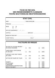
Sheet of clinical information
Informations for the patients<br />
Professeur Didier RAOULT<br />
HOPITAL DE LA TIMONE<br />
Phone. 33. 4.91.38.55.14/19<br />
Mail : didier.raoult@medecine.univ-mrs.fr<br />
The Whipple’s <strong>disease</strong> is caused by a bacterium called <strong>Tropheryma</strong><br />
<strong>whipplei</strong>. The bacterium lives probably in the environment, maybe water,<br />
but we do not really know the source of the infection. The <strong>disease</strong> seems<br />
to be more frequent in some geographical area, mainly, for France in<br />
Rhône-Alpes area, but also in Switzerl<strong>and</strong> <strong>and</strong> Germany.<br />
The presentations of the <strong>disease</strong> are diverse. The most frequent<br />
<strong>and</strong> classical form is those with digestive signs including a chronic<br />
diarrhea, associated with a weight loss <strong>and</strong> a skin which become more<br />
dark. This form is often associated to the presence of adenopathies or<br />
arthralgia, which could be the first clinical signs of the <strong>disease</strong>.<br />
Sometimes, this <strong>disease</strong> is misdiagnosed with rheumatoid polyarthritis.<br />
In the classical from, the localization of the bacterium on the small-bowel<br />
is systematic. This fact explains why we perform a digestive fibroscopy<br />
with a biopsy to check for the presence of the bacterium at the beginning<br />
<strong>and</strong> in the follow-up of the <strong>disease</strong>. Besides, the bacterium could also<br />
infects different sites such as the brain or the cardiac valves.<br />
Without treatment, the evolution of Whipple’s <strong>disease</strong> is always<br />
fatale. The treatment is long (always superior to 1 year) <strong>and</strong> after the end<br />
of the treatment, sometimes even after very long treatment, relapses are<br />
possible, mainly in the brain. A regular follow-up of patients with<br />
Whipple’s <strong>disease</strong> is thus necessary.<br />
The bacterium has been cultivated for the first time in our<br />
laboratory in Marseille (France), in 1999, allowing new perspectives for<br />
the diagnosis <strong>and</strong> the management of this <strong>disease</strong>.
The diagnosis could be performed by the detection of the<br />
bacterium or its genes, by molecular techniques. We look for the<br />
presence of the bacterium in blood, saliva <strong>and</strong> stools, for all the patients.<br />
For the patients with a digestive localization, we look for the presence of<br />
the bacterium in the small-bowel digestive, one time per year. For the<br />
patients who present a brain localization, we look for the bacterium on<br />
cerebrospinal fluid obtained by lumbar puncture. The lumbar puncture is<br />
first performed for the diagnosis, <strong>and</strong> 2 months after the beginning of the<br />
treatment, to check the disappearance of the bacterium. After the end of<br />
the treatment, a lumber puncture should be proposed annually, to allow<br />
the detection of a relapse. Some radiological brain exploration could be<br />
also justified in this context.<br />
For the treatment, the knowledge has progressed quickly these<br />
last years, with the culture of the bacterium. For classic Whipple’s<br />
<strong>disease</strong>, it seems that the treatment the more efficiency is the<br />
association of doxycycline <strong>and</strong> hydroxychloroquine. Hydroxychloroquine<br />
allows doxycycline to be efficient in the cells. For the neurological form,<br />
the reference treatment includes cotrimoxazole. Unfortunately, relapses<br />
have been reported even with these approaches. The treatment should<br />
never been less than 1 year, <strong>and</strong> after a follow-up during all the life is<br />
required to detect a relapse.<br />
The results of your sampling, for the diagnosis, the treatment <strong>and</strong><br />
the follow-up (whatever the documentation) are susceptible to be used<br />
(anonymously) for reports about your <strong>disease</strong> in the context of a<br />
scientific publication.


