

Development of the optic nerve of the rat - Investigative ...
Development of the optic nerve of the rat - Investigative ...
Development of the optic nerve of the rat - Investigative ...

Create successful ePaper yourself
Turn your PDF publications into a flip-book with our unique Google optimized e-Paper software.
Volume 14<br />
Number 10<br />
Rat <strong>optic</strong> <strong>nerve</strong> 741<br />
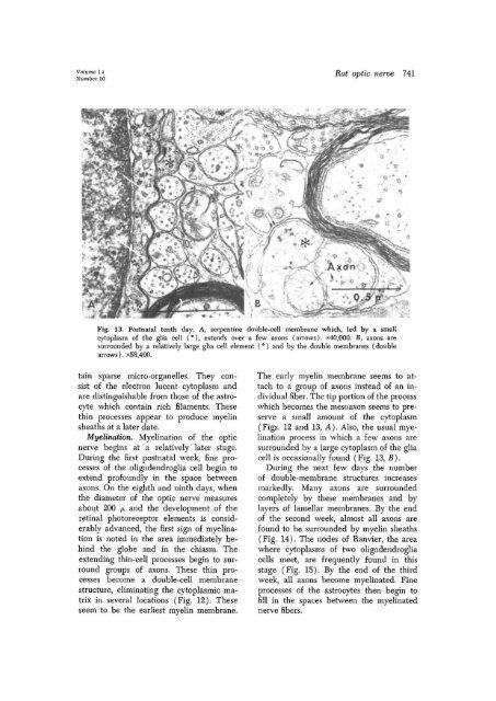
Fig. 13. Postnatal tenth day. A, serpentine double-cell membrane which, led by a small<br />
cytoplasm <strong>of</strong> <strong>the</strong> glia cell (°), extends over a few axons (arrows). x40,000. B> axons are<br />
surrounded by a relatively large glia cell element (*) and by <strong>the</strong> double membranes (double<br />
arrows). x58,400.<br />
tain sparse micro-organelles. They consist<br />
<strong>of</strong> <strong>the</strong> electron lucent cytoplasm and<br />
are distinguishable from those <strong>of</strong> <strong>the</strong> astrocyte<br />
which contain rich filaments. These<br />
thin processes appear to produce myelin<br />
sheaths at a later date,<br />
Myelination. Myelination <strong>of</strong> <strong>the</strong> <strong>optic</strong><br />
<strong>nerve</strong> begins at a relatively later stage.<br />
During <strong>the</strong> first postnatal week, fine processes<br />
<strong>of</strong> <strong>the</strong> oligodendroglia cell begin to<br />
extend pr<strong>of</strong>oundly in <strong>the</strong> space between<br />
axons. On <strong>the</strong> eighth and ninth days, when<br />
<strong>the</strong> diameter <strong>of</strong> <strong>the</strong> <strong>optic</strong> <strong>nerve</strong> measures<br />
about 200 ju and <strong>the</strong> development <strong>of</strong> <strong>the</strong><br />
retinal photoreceptor elements is considerably<br />
advanced, <strong>the</strong> first sign <strong>of</strong> myelination<br />
is noted in <strong>the</strong> area immediately behind<br />
<strong>the</strong> globe and in <strong>the</strong> chiasm. The<br />
extending thin-cell processes begin to surround<br />
groups <strong>of</strong> axons. These thin processes<br />
become a double-cell membrane<br />
structure, eliminating <strong>the</strong> cytoplasmic matrix<br />
in several locations (Fig. 12). These<br />
seem to be <strong>the</strong> earliest myelin membrane.<br />
The early myelin membrane seems to attach<br />
to a group <strong>of</strong> axons instead <strong>of</strong> an individual<br />
fiber. The tip portion <strong>of</strong> <strong>the</strong> process<br />
which becomes <strong>the</strong> mesoaxon seems to preserve<br />
a small amount <strong>of</strong> <strong>the</strong> cytoplasm<br />
(Figs. 12 and 13, A). Also, <strong>the</strong> usual myelination<br />
process in which a few axons are<br />
surrounded by a large cytoplasm <strong>of</strong> <strong>the</strong> glia<br />
cell is occasionally found (Fig. 13, B).<br />
During <strong>the</strong> next few days <strong>the</strong> number<br />
<strong>of</strong> double-membrane structures increases<br />
markedly. Many axons are surrounded<br />
completely by <strong>the</strong>se membranes and by<br />
layers <strong>of</strong> lamellar membranes. By <strong>the</strong> end<br />
<strong>of</strong> <strong>the</strong> second week, almost all axons are<br />
found to be surrounded by myelin sheaths<br />
(Fig. 14). The nodes <strong>of</strong> Ranvier, <strong>the</strong> area<br />
where cytoplasms <strong>of</strong> two oligodendroglia<br />
cells meet, are frequently found in this<br />
stage (Fig. 15). By <strong>the</strong> end <strong>of</strong> <strong>the</strong> third<br />
week, all axons become myelinated. Fine<br />
processes <strong>of</strong> <strong>the</strong> astrocytes <strong>the</strong>n begin to<br />
fill in <strong>the</strong> spaces between <strong>the</strong> myelinated<br />
<strong>nerve</strong> fibers.


