

Development of the optic nerve of the rat - Investigative ...
Development of the optic nerve of the rat - Investigative ...
Development of the optic nerve of the rat - Investigative ...

You also want an ePaper? Increase the reach of your titles
YUMPU automatically turns print PDFs into web optimized ePapers that Google loves.
742 Kuioabara <strong>Investigative</strong> Ophthalmology<br />
October 1975<br />
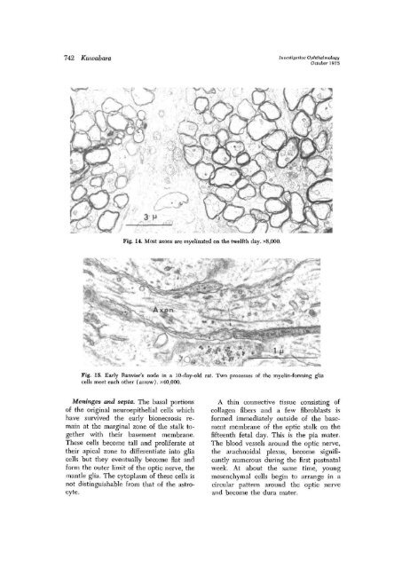
Fig. 14. Most axons are myelinated on <strong>the</strong> twelfth day. x8,000.<br />
O*e=»»<br />
Fig. 15. Early Ranvier's node in a 10-day-old <strong>rat</strong>. Two processes <strong>of</strong> <strong>the</strong> myelin-fomiing glia<br />
cells meet each o<strong>the</strong>r (arrow). x40 ; 000.<br />
Meninges and septa. The basal portions<br />
<strong>of</strong> <strong>the</strong> original neuroepi<strong>the</strong>lial cells which<br />
have survived <strong>the</strong> early bionecrosis remain<br />
at <strong>the</strong> marginal zone <strong>of</strong> <strong>the</strong> stalk toge<strong>the</strong>r<br />
with <strong>the</strong>ir basement membrane.<br />
These cells become tall and prolife<strong>rat</strong>e at<br />
<strong>the</strong>ir apical zone to differentiate into glia<br />
cells but <strong>the</strong>y eventually become flat and<br />
form <strong>the</strong> outer limit <strong>of</strong> <strong>the</strong> <strong>optic</strong> <strong>nerve</strong>, <strong>the</strong><br />
mantle glia. The cytoplasm <strong>of</strong> <strong>the</strong>se cells is<br />
not distinguishable from that <strong>of</strong> <strong>the</strong> astrocyte.<br />
A thin connective tissue consisting <strong>of</strong><br />
collagen fibers and a few fibroblasts is<br />
formed immediately outside <strong>of</strong> <strong>the</strong> basement<br />
membrane <strong>of</strong> <strong>the</strong> <strong>optic</strong> stalk on <strong>the</strong><br />
fifteenth fetal day. This is <strong>the</strong> pia mater.<br />
The blood vessels around <strong>the</strong> <strong>optic</strong> <strong>nerve</strong>,<br />
<strong>the</strong> arachnoidal plexus, become significantly<br />
numerous during <strong>the</strong> first postnatal<br />
week. At about <strong>the</strong> same time, young<br />
mesenchymal cells begin to arrange in a<br />
circular pattern around <strong>the</strong> <strong>optic</strong> <strong>nerve</strong><br />
and become <strong>the</strong> dura mater.


