

Development of the optic nerve of the rat - Investigative ...
Development of the optic nerve of the rat - Investigative ...
Development of the optic nerve of the rat - Investigative ...

Create successful ePaper yourself
Turn your PDF publications into a flip-book with our unique Google optimized e-Paper software.
Volume 14<br />
Number 10<br />
Rat <strong>optic</strong> <strong>nerve</strong> 735<br />
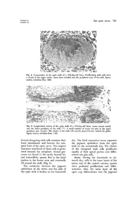
Fig. 4. Cross-section <strong>of</strong> <strong>the</strong> <strong>optic</strong> stalk <strong>of</strong> a 15.5-day-old fetus. Prolife<strong>rat</strong>ing stalk cells form<br />
a cluster in <strong>the</strong> upper center. Axons have invaded into <strong>the</strong> peripheral zone <strong>of</strong> <strong>the</strong> stalk. Epoxy<br />
section, toluidine blue. *260.<br />
Fig. 5. Longitudinal section <strong>of</strong> <strong>the</strong> <strong>optic</strong> stalk <strong>of</strong> a 15.5-day-old fetus. Axons extend mainly<br />
into <strong>the</strong> lower periphery <strong>of</strong> <strong>the</strong> stalk ( *), A small number <strong>of</strong> axons are seen in <strong>the</strong> upper<br />
periphery also (arrow). The cluster <strong>of</strong> <strong>the</strong> stalk cell extends about 0.5 mm. behind <strong>the</strong> globe.<br />
Epoxy section, toluidine blue. *180.<br />
Several elongating stalk cells maintain <strong>the</strong>ir<br />
basal attachments and become <strong>the</strong> marginal<br />
limit <strong>of</strong> <strong>the</strong> <strong>optic</strong> <strong>nerve</strong>. The original<br />
basement membrane <strong>of</strong> <strong>the</strong>se cells is prominent<br />
beneath <strong>the</strong> cytoplasm. Axonal processes<br />
are found in <strong>the</strong> newly formed lateral<br />
intercellular spaces, first in <strong>the</strong> basal<br />
portion in <strong>the</strong> fissure zone and eventually<br />
all around <strong>the</strong> stalk (Fig. 5).<br />
The continuity between <strong>the</strong> pigment<br />
epi<strong>the</strong>lium <strong>of</strong> <strong>the</strong> retina and <strong>the</strong> cells <strong>of</strong><br />
<strong>the</strong> <strong>optic</strong> stalk is broken on <strong>the</strong> fourteenth<br />
day. The thick connective tissue sepa<strong>rat</strong>es<br />
<strong>the</strong> pigment epi<strong>the</strong>lium from <strong>the</strong> <strong>optic</strong><br />
stalk on <strong>the</strong> seventeenth day. The cluster<br />
<strong>of</strong> <strong>the</strong> elongated stalk cells prolife<strong>rat</strong>e<br />
rapidly at <strong>the</strong>ir apical portion and differentiate<br />
into glia cells.<br />
Axons. During <strong>the</strong> fourteenth to sixteenth<br />
day, cells in <strong>the</strong> inner layers <strong>of</strong> <strong>the</strong><br />
retina and <strong>of</strong> <strong>the</strong> central nervous system<br />
show pr<strong>of</strong>ound prolife<strong>rat</strong>ion and differentiation.<br />
Also, <strong>the</strong> outer layer <strong>of</strong> <strong>the</strong><br />
<strong>optic</strong> cup differentiates into <strong>the</strong> pigment


