

Development of the optic nerve of the rat - Investigative ...
Development of the optic nerve of the rat - Investigative ...
Development of the optic nerve of the rat - Investigative ...

You also want an ePaper? Increase the reach of your titles
YUMPU automatically turns print PDFs into web optimized ePapers that Google loves.
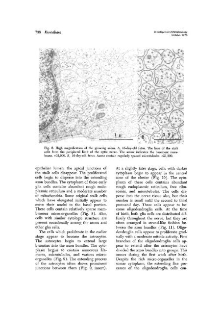
738 Kuwabara Investigntive Ophthalmology<br />
October 1975<br />
Fig. 8. High magnification <strong>of</strong> <strong>the</strong> growing axons. A, lS-day-old fetus. The base <strong>of</strong> <strong>the</strong> stalk<br />
cells form <strong>the</strong> peripheral limit <strong>of</strong> <strong>the</strong> <strong>optic</strong> <strong>nerve</strong>. The arrow indicates <strong>the</strong> basement membrane.<br />
x32,000. B, 16-day-old fetus. Axons contain regularly spaced microtubules. x51,200.<br />
epi<strong>the</strong>liar lumen, <strong>the</strong> apical junctions <strong>of</strong><br />
<strong>the</strong> stalk cells disappear. The prolife<strong>rat</strong>ed<br />
cells begin to disperse into <strong>the</strong> extending<br />
axon bundles. The cytoplasm <strong>of</strong> <strong>the</strong>se early<br />
glia cells contains abundant rough endoplasmic<br />
reticulum and a mode<strong>rat</strong>e number<br />
<strong>of</strong> mitochondria. Some original stalk cells<br />
which have elongated initially appear to<br />
move <strong>the</strong>ir nuclei to <strong>the</strong> basal portion.<br />
These cells contain relatively sparse membranous<br />
micro-organelles (Fig. 8). Also,<br />
cells with similar cytologic structure are<br />
present occasionally among <strong>the</strong> axons and<br />
o<strong>the</strong>r glia cells.<br />
The cells which prolife<strong>rat</strong>e in <strong>the</strong> earlier<br />
stage appear to become <strong>the</strong> astrocytes.<br />
The astrocytes begin to extend large<br />
branches into <strong>the</strong> axon bundles. The cytoplasm<br />
begins to contain numerous filaments,<br />
microtubules, and various microorganelles<br />
(Fig. 9). The extending process<br />
<strong>of</strong> <strong>the</strong> astrocytes <strong>of</strong>ten shows prominent<br />
junctions between <strong>the</strong>m (Fig. 9, insert).<br />
At a slightly later stage, cells with darker<br />
cytoplasm begin to appear in <strong>the</strong> central<br />
zone <strong>of</strong> <strong>the</strong> cluster (Fig. 10). The cytoplasm<br />
<strong>of</strong> <strong>the</strong>se cells contains abundant<br />
rough endoplasmic reticulum, free ribosomes,<br />
and microtubules. The cells disperse<br />
into <strong>the</strong> <strong>nerve</strong> tissue also, but <strong>the</strong>ir<br />
number is small until <strong>the</strong> second to third<br />
postnatal day. These cells appear to become<br />
oligodendroglia cells. At <strong>the</strong> time<br />
<strong>of</strong> birth, both glia cells are distributed diffusely<br />
throughout <strong>the</strong> <strong>nerve</strong>, but <strong>the</strong>y are<br />
<strong>of</strong>ten arranged in strand-like fashion between<br />
<strong>the</strong> axon bundles (Fig. 11). Oligodendroglia<br />
cells appear to prolife<strong>rat</strong>e gradually<br />
with a mode<strong>rat</strong>e mitotic activity. Fine<br />
branches <strong>of</strong> <strong>the</strong> oligodendroglia cells appear<br />
to extend after <strong>the</strong> astrocytes have<br />
divided <strong>the</strong> axon bundles into groups. This<br />
occurs during <strong>the</strong> first week after birth.<br />
Despite <strong>the</strong> rich micro-organelles in <strong>the</strong><br />
somar cytoplasm, <strong>the</strong> extending fine processes<br />
<strong>of</strong> <strong>the</strong> oligodendroglia cells con-


