

Development of the optic nerve of the rat - Investigative ...
Development of the optic nerve of the rat - Investigative ...
Development of the optic nerve of the rat - Investigative ...

You also want an ePaper? Increase the reach of your titles
YUMPU automatically turns print PDFs into web optimized ePapers that Google loves.
734 Kuioabara <strong>Investigative</strong> Ophthalmology<br />
October 1975<br />
•'.<br />
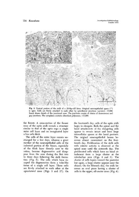
Fig. 3. Central portion <strong>of</strong> <strong>the</strong> stalk <strong>of</strong> a 15-day-old fetus. Original neuroepi<strong>the</strong>lial space (*)<br />
is open. Cells are firmly attached to each o<strong>the</strong>r by apicolateral junctions (arrows). x7,000.<br />
Insert shows details <strong>of</strong> <strong>the</strong> junctional zone. The junctions consist <strong>of</strong> chains <strong>of</strong> desmosomes and<br />
gap junctions, The cytoplasm contains abundant polysonies. xl7,000.<br />
<strong>the</strong> fissure. A cross-section <strong>of</strong> <strong>the</strong> fissure<br />
zone <strong>of</strong> <strong>the</strong> <strong>optic</strong> stalk reveals a structure<br />
similar to that <strong>of</strong> <strong>the</strong> <strong>optic</strong> cup—a single<br />
outer cell layer and an invaginated layer<br />
a few cells thick.<br />
The cells <strong>of</strong> <strong>the</strong> outer layer remain unchanged<br />
for a few days, whereas a great<br />
number <strong>of</strong> <strong>the</strong> neuroepi<strong>the</strong>lial cells <strong>of</strong> <strong>the</strong><br />
indented portion <strong>of</strong> <strong>the</strong> fissure, especially<br />
<strong>of</strong> <strong>the</strong> thick layer directly next to <strong>the</strong><br />
retina, become degene<strong>rat</strong>ive and disappear<br />
from <strong>the</strong> area during <strong>the</strong> first two<br />
to three days following <strong>the</strong> stalk formation<br />
(Fig. 2). The cells which have escaped<br />
<strong>the</strong> degene<strong>rat</strong>ion form a tube-like<br />
tissue <strong>of</strong> a single cell layer. These cells<br />
are firmly attached to each o<strong>the</strong>r at <strong>the</strong><br />
apicolateral zone (Figs. 3 and 17). On<br />
<strong>the</strong> fourteenth day, cells <strong>of</strong> <strong>the</strong> <strong>optic</strong> stalk<br />
begin to elongate. Both <strong>the</strong> apical and <strong>the</strong><br />
basal attachments <strong>of</strong> <strong>the</strong> elongating cells<br />
appear to remain intact and form large<br />
intercellular spaces at <strong>the</strong> basal portions.<br />
The original neuroepi<strong>the</strong>lial lumen becomes<br />
almost nonexistent on <strong>the</strong> fourteenth<br />
day. Prolife<strong>rat</strong>ion <strong>of</strong> <strong>the</strong> stalk cells<br />
with mitotic activity is observed at <strong>the</strong><br />
apical zone until <strong>the</strong> sixteenth day. The<br />
prolife<strong>rat</strong>ed cells which have no basal attachment<br />
form a large cluster at <strong>the</strong><br />
retrobulbar area (Figs. 4 and 5). The<br />
cluster <strong>of</strong> cells tapers toward <strong>the</strong> posterior<br />
but again, a large cluster appears near <strong>the</strong><br />
chiasm. On <strong>the</strong> fifteenth day, <strong>the</strong> stalk becomes<br />
an oval piece having a cluster <strong>of</strong><br />
cells in <strong>the</strong> upper, <strong>of</strong>f-center zone (Fig. 4).


