

Development of the optic nerve of the rat - Investigative ...
Development of the optic nerve of the rat - Investigative ...
Development of the optic nerve of the rat - Investigative ...

Create successful ePaper yourself
Turn your PDF publications into a flip-book with our unique Google optimized e-Paper software.
Volume 14<br />
Number 10<br />
Rat <strong>optic</strong> <strong>nerve</strong> 743<br />
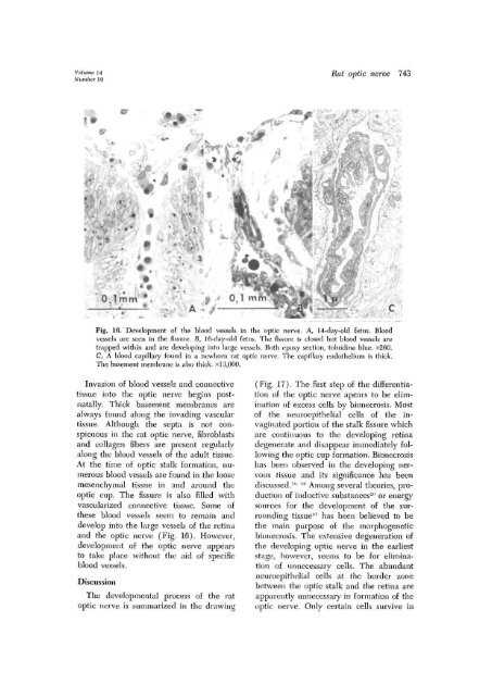
Fig. 16. <strong>Development</strong> <strong>of</strong> <strong>the</strong> blood vessels in <strong>the</strong> <strong>optic</strong> <strong>nerve</strong>. A, 14-day-old fetus. Blood<br />
vessels are seen in <strong>the</strong> fissure. B, lG-day-old fetus. The fissure is closed but blood vessels are<br />
trapped within and are developing into large vessels. Both epoxy section, toluidine blue. x260.<br />
C, A blood capillary found in a newborn <strong>rat</strong> <strong>optic</strong> <strong>nerve</strong>. The capillary endo<strong>the</strong>lium is thick-.<br />
The basement membrane is also thick, x 13,000.<br />
Invasion <strong>of</strong> blood vessels and connective<br />
tissue into <strong>the</strong> <strong>optic</strong> <strong>nerve</strong> begins postnatally.<br />
Thick basement membranes are<br />
always found along <strong>the</strong> invading vascular<br />
tissue. Although <strong>the</strong> septa is not conspicuous<br />
in <strong>the</strong> <strong>rat</strong> <strong>optic</strong> <strong>nerve</strong>, fibroblasts<br />
and collagen fibers are present regularly<br />
along <strong>the</strong> blood vessels <strong>of</strong> <strong>the</strong> adult tissue.<br />
At <strong>the</strong> time <strong>of</strong> <strong>optic</strong> stalk formation, numerous<br />
blood vessels are found in <strong>the</strong> loose<br />
mesenchymal tissue in and around <strong>the</strong><br />
<strong>optic</strong> cup. The fissure is also filled with<br />
vascularized connective tissue. Some <strong>of</strong><br />
<strong>the</strong>se blood vessels seem to remain and<br />
develop into <strong>the</strong> large vessels <strong>of</strong> <strong>the</strong> retina<br />
and <strong>the</strong> <strong>optic</strong> <strong>nerve</strong> (Fig. 16). However,<br />
development <strong>of</strong> <strong>the</strong> <strong>optic</strong> <strong>nerve</strong> appears<br />
to take place without <strong>the</strong> aid <strong>of</strong> specific<br />
blood vessels.<br />
Discussion<br />
The developmental process <strong>of</strong> <strong>the</strong> <strong>rat</strong><br />
<strong>optic</strong> <strong>nerve</strong> is summarized in <strong>the</strong> drawing<br />
(Fig. 17). The first step <strong>of</strong> <strong>the</strong> differentiation<br />
<strong>of</strong> <strong>the</strong> <strong>optic</strong> <strong>nerve</strong> apears to be elimination<br />
<strong>of</strong> excess cells by bionecrosis. Most<br />
<strong>of</strong> <strong>the</strong> neuroepi<strong>the</strong>lial cells <strong>of</strong> <strong>the</strong> invaginated<br />
portion <strong>of</strong> <strong>the</strong> stalk fissure which<br />
are continuous to <strong>the</strong> developing retina<br />
degene<strong>rat</strong>e and disappear immediately following<br />
<strong>the</strong> <strong>optic</strong> cup formation. Bionecrosis<br />
has been observed in <strong>the</strong> developing nervous<br />
tissue and its significance has been<br />
discussed. IS - l!1 Among several <strong>the</strong>ories, production<br />
<strong>of</strong> inductive substances-" or energy<br />
sources for <strong>the</strong> development <strong>of</strong> <strong>the</strong> surrounding<br />
tissue'-' 1 has been believed to be<br />
<strong>the</strong> main purpose <strong>of</strong> <strong>the</strong> morphogenetic<br />
bionecrosis. The extensive degene<strong>rat</strong>ion <strong>of</strong><br />
<strong>the</strong> developing <strong>optic</strong> <strong>nerve</strong> in <strong>the</strong> earliest<br />
stage, however, seems to be for elimination<br />
<strong>of</strong> unnecessary cells. The abundant<br />
neuroepi<strong>the</strong>lial cells at <strong>the</strong> border zone<br />
between <strong>the</strong> <strong>optic</strong> stalk and <strong>the</strong> retina are<br />
apparently unnecessary in formation <strong>of</strong> <strong>the</strong><br />
<strong>optic</strong> <strong>nerve</strong>. Only certain cells survive in


