

Development of the optic nerve of the rat - Investigative ...
Development of the optic nerve of the rat - Investigative ...
Development of the optic nerve of the rat - Investigative ...

Create successful ePaper yourself
Turn your PDF publications into a flip-book with our unique Google optimized e-Paper software.
Volume 14<br />
Number 10<br />
Rat <strong>optic</strong> <strong>nerve</strong> 733<br />
brain tissue was processed as one piece in early<br />
fetuses, and <strong>the</strong> <strong>optic</strong> <strong>nerve</strong>s <strong>of</strong> animals older<br />
than <strong>the</strong> eighteenth embryonal day were divided<br />
into three pieces: postbulbar, central (midcanalicular),<br />
and chiasma portions. The tissue<br />
was postfixed in 1 per cent osmium tetroxide in<br />
<strong>the</strong> same buffer solution for 90 minutes at 4° C.<br />
After fur<strong>the</strong>r trimming and orientation under a<br />
dissecting microscope, <strong>the</strong> small pieces were dehyd<strong>rat</strong>ed<br />
in a series <strong>of</strong> ethyl alcohol, treated with<br />
propylene oxide, and embedded in an epoxy resin<br />
following Luft's method. 1 "<br />
Sections (0.5 to 1.0 y. thick) were stained with<br />
alkaline toluidine blue for light microscopic study.<br />
Ult<strong>rat</strong>hin sections were stained with uranyl acetate<br />
and lead cit<strong>rat</strong>e and were examined by an electron<br />
microscope with an accele<strong>rat</strong>ing voltage <strong>of</strong><br />
80 KV.<br />
Results<br />
The <strong>optic</strong> stalk, measuring about 200 /± in<br />
length, becomes recognizable immediately<br />
following <strong>the</strong> formation <strong>of</strong> <strong>the</strong> <strong>optic</strong> cup<br />
on <strong>the</strong> thirteenth fetal day (Fig. 1). The<br />
invaginating fold <strong>of</strong> <strong>the</strong> inner layer <strong>of</strong> <strong>the</strong><br />
cup extends posteriorly and becomes <strong>the</strong><br />
fissure which disappears rapidly as development<br />
progresses. The whitish <strong>nerve</strong><br />
tissue becomes grossly visible on about <strong>the</strong><br />
sixteenth day. The most active differentiation<br />
<strong>of</strong> <strong>the</strong> <strong>optic</strong> <strong>nerve</strong> appears to take<br />
place between <strong>the</strong> sixteenth and eighteenth<br />
day.<br />
On <strong>the</strong> day <strong>of</strong> birth, <strong>the</strong> <strong>optic</strong> <strong>nerve</strong> is<br />
about 2 mm. long and <strong>the</strong> configu<strong>rat</strong>ion<br />
<strong>of</strong> <strong>the</strong> chiasma is clearly formed. Myelination<br />
<strong>of</strong> <strong>the</strong> <strong>nerve</strong> fibers, however, begins<br />
on <strong>the</strong> postnatal fifth day and continues<br />
actively until <strong>the</strong> end <strong>of</strong> <strong>the</strong> second week.<br />
The developmental process appears to<br />
slow down considerably by <strong>the</strong> end <strong>of</strong> <strong>the</strong><br />
third postnatal week and no appreciable<br />
alte<strong>rat</strong>ion in <strong>the</strong> cytologic appearance is<br />
observed after this period.<br />
Optic stalk. The tubular <strong>optic</strong> stalk is<br />
formed at <strong>the</strong> isthmus <strong>of</strong> <strong>the</strong> neurovesicle<br />
between <strong>the</strong> outpouching ocular tissue and<br />
<strong>the</strong> central nervous system. The earliest<br />
<strong>optic</strong> stalk consists <strong>of</strong> a single layer <strong>of</strong><br />
neuroepi<strong>the</strong>lial cells. The invagination <strong>of</strong><br />
<strong>the</strong> <strong>optic</strong> cup extends posteriorly into <strong>the</strong><br />
lower portion <strong>of</strong> <strong>the</strong> stalk and becomes<br />
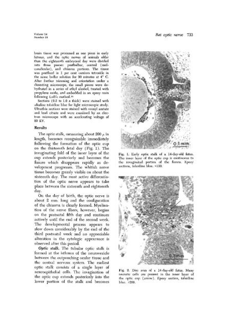
Fig. 1. Early <strong>optic</strong> stalk <strong>of</strong> a 14-day-old fetus.<br />
The inner layer <strong>of</strong> <strong>the</strong> <strong>optic</strong> cup is continuous to<br />
<strong>the</strong> invaginated portion <strong>of</strong> <strong>the</strong> fissure. Epoxy<br />
sections, toluidine blue. xl30.<br />
Fig. 2. Disc area <strong>of</strong> a 14-day-old fetus. Many<br />
necrotic cells are present in <strong>the</strong> inner layer <strong>of</strong><br />
<strong>the</strong> <strong>optic</strong> cup (arrow). Epoxy section, toluidine<br />
blue. x260.


