

Development of the optic nerve of the rat - Investigative ...
Development of the optic nerve of the rat - Investigative ...
Development of the optic nerve of the rat - Investigative ...

You also want an ePaper? Increase the reach of your titles
YUMPU automatically turns print PDFs into web optimized ePapers that Google loves.
Volume 14<br />
Number 10<br />
Rat <strong>optic</strong> <strong>nerve</strong> 737<br />
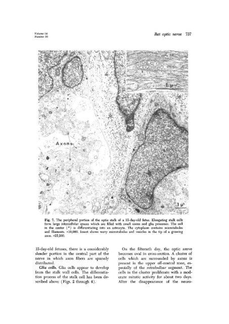
Fig. 7. The peripheral portion <strong>of</strong> <strong>the</strong> <strong>optic</strong> stalk <strong>of</strong> a 15-day-old fetus. Elongating stalk cells<br />
form large intercellular spaces which are filled with small axons and glia processes. The cell<br />
in <strong>the</strong> center (*) is differentiating into an astrocyte. The cytoplasm contains microtubules<br />
and filaments. xl0,000. Insert shows wavy microtubules and vesicles in <strong>the</strong> tip <strong>of</strong> a growing<br />
axon. x25,200.<br />
15-day-old fetuses, <strong>the</strong>re is a considerably<br />
slender portion in <strong>the</strong> central part <strong>of</strong> <strong>the</strong><br />
<strong>nerve</strong> in which axon fibers are sparsely<br />
distributed.<br />
Glia cells. Glia cells appear to develop<br />
from <strong>the</strong> stalk wall cells. The differentiation<br />
process <strong>of</strong> <strong>the</strong> stalk cell has been described<br />
above (Figs. 2 through 4).<br />
On <strong>the</strong> fifteenth day, <strong>the</strong> <strong>optic</strong> <strong>nerve</strong><br />
becomes oval in cross-section. A cluster <strong>of</strong><br />
cells which are surrounded by axons is<br />
present in <strong>the</strong> upper <strong>of</strong>f-central zone, especially<br />
<strong>of</strong> <strong>the</strong> retrobulbar segment. The<br />
cells in <strong>the</strong> cluster prolife<strong>rat</strong>e with a mode<strong>rat</strong>e<br />
mitotic activity for about two days.<br />
After <strong>the</strong> disappearance <strong>of</strong> <strong>the</strong> neuro-


