Granulocyte colony-stimulating factor (G-CSF ... - ResearchGate
Granulocyte colony-stimulating factor (G-CSF ... - ResearchGate
Granulocyte colony-stimulating factor (G-CSF ... - ResearchGate

You also want an ePaper? Increase the reach of your titles
YUMPU automatically turns print PDFs into web optimized ePapers that Google loves.
224 BRAIN RESEARCH 1149 (2007) 223– 231<br />
2003), are candidates for cell therapy of spinal cord injury,<br />
because they can be transplanted autologously. Indeed,<br />
transplantation of HSC promotes functional recovery after<br />
compression-induced spinal cord injury in mice, as we<br />
reported previously (Koshizuka et al., 2004; Koda et al., 2005),<br />
and transplantation of BMSC significantly improves hind limb<br />
function after spinal cord injury in mice and rats (Chopp et al.,<br />
2000; Hofstetter et al., 2002; Wu et al., 2003). These lines of<br />
evidence show the potential of bone marrow-derived stem<br />
cells to restore injured spinal cord tissue and to promote<br />
functional recovery.<br />
<strong>Granulocyte</strong> <strong>colony</strong>-<strong>stimulating</strong> <strong>factor</strong> (G-<strong>CSF</strong>) is widely<br />
known as a cytokine that induces survival, proliferation and<br />
differentiation of cells of the neutrophil lineage (Nicola et<br />
al., 1983; Roberts, 2005). Further, G-<strong>CSF</strong> can mobilize bone<br />
marrow cells into peripheral blood, an action used clinically<br />
for patients with leukocytopenia and for donors of peripheral<br />
blood-derived HSCs for transplantation (Jansen et al.,<br />
2005). Bone marrow-derived stem cells mobilized by G-<strong>CSF</strong><br />
may have the potential to migrate into and repair various<br />
injured tissues. For example, G-<strong>CSF</strong> has been shown to<br />
mobilize bone marrow-derived stem cells to repair the<br />
ischemic myocardium (Orlic et al., 2001; Kawada et al.,<br />
2004). As for the central nervous system, bone marrow cells<br />
mobilized by G-<strong>CSF</strong> migrate into, survive and express a<br />
neural phenotype in normal brain (Corti et al., 2002a), spinal<br />
cord and dorsal root ganglia (Corti et al., 2002b). Moreover,<br />
G-<strong>CSF</strong> mobilized bone marrow cells into the ischemic brain<br />
and promoted functional recovery (Kawada et al., 2006). The<br />
influence of G-<strong>CSF</strong>-mediated mobilization of bone marrowderived<br />
stem cells on the injured spinal cord remains to be<br />
elucidated.<br />
Here we showed that G-<strong>CSF</strong> promoted the mobilization and<br />
migration of bone marrow cells into the spinal cord, the<br />
restoration of damaged spinal cord tissue and the recovery of<br />
hind limb function.<br />
2. Results<br />
Mice tolerated the irradiation and bone marrow transplantation<br />
well. FACS analysis showed that approximately 80% of the<br />
whole bone marrow cells were positive for GFP 4 weeks after<br />
bone marrow transplantation, indicating that GFP Tg-derived<br />
bone marrow cells survived and reconstituted hematopoiesis<br />
in grafted mice (not shown).<br />
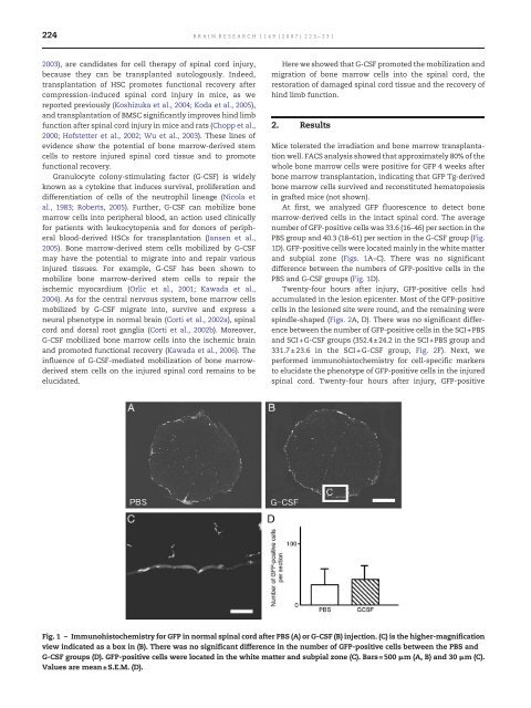
At first, we analyzed GFP fluorescence to detect bone<br />
marrow-derived cells in the intact spinal cord. The average<br />
number of GFP-positive cells was 33.6 (16–46) per section in the<br />
PBS group and 40.3 (18–61) per section in the G-<strong>CSF</strong> group (Fig.<br />
1D). GFP-positive cells were located mainly in the white matter<br />
and subpial zone (Figs. 1A–C). There was no significant<br />
difference between the numbers of GFP-positive cells in the<br />
PBS and G-<strong>CSF</strong> groups (Fig. 1D).<br />
Twenty-four hours after injury, GFP-positive cells had<br />
accumulated in the lesion epicenter. Most of the GFP-positive<br />
cells in the lesioned site were round, and the remaining were<br />
spindle-shaped (Figs. 2A, D). There was no significant difference<br />
between the number of GFP-positive cells in the SCI+PBS<br />
and SCI+G-<strong>CSF</strong> groups (352.4±24.2 in the SCI+PBS group and<br />
331.7±23.6 in the SCI+G-<strong>CSF</strong> group, Fig. 2F). Next, we<br />
performed immunohistochemistry for cell-specific markers<br />
to elucidate the phenotype of GFP-positive cells in the injured<br />
spinal cord. Twenty-four hours after injury, GFP-positive<br />
Fig. 1 – Immunohistochemistry for GFP in normal spinal cord after PBS (A) or G-<strong>CSF</strong> (B) injection. (C) is the higher-magnification<br />
view indicated as a box in (B). There was no significant difference in the number of GFP-positive cells between the PBS and<br />
G-<strong>CSF</strong> groups (D). GFP-positive cells were located in the white matter and subpial zone (C). Bars=500 μm (A, B) and 30 μm (C).<br />
Values are mean±S.E.M. (D).