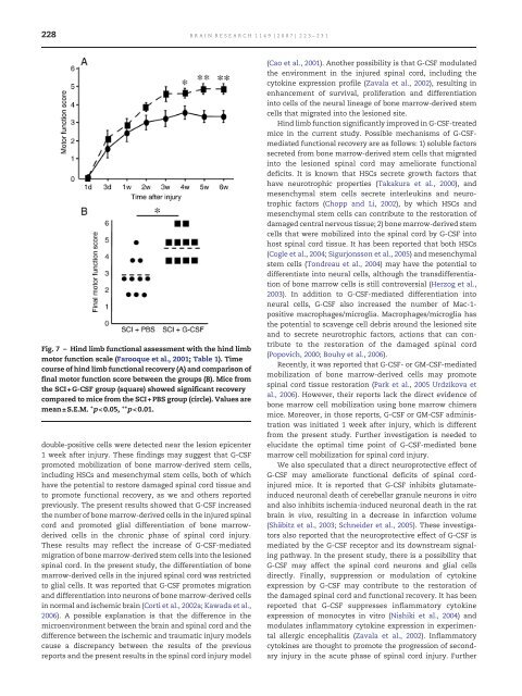
228 BRAIN RESEARCH 1149 (2007) 223– 231 Fig. 7 – Hind limb functional assessment with the hind limb motor function scale (Farooque et al., 2001; Table 1). Time course of hind limb functional recovery (A) and comparison of final motor function score between the groups (B). Mice from the SCI+G-<strong>CSF</strong> group (square) showed significant recovery compared to mice from the SCI+PBS group (circle). Values are mean± S.E.M. ⁎p
BRAIN RESEARCH 1149 (2007) 223– 231 229 investigation is needed to elucidate these mechanisms of action of G-<strong>CSF</strong>. Regarding the clinical application of G-<strong>CSF</strong> for the treatment of spinal cord injury, G-<strong>CSF</strong> has some advantages over other treatments under investigation. First, G-<strong>CSF</strong> has already been approved as a safe drug, and it is widely used in clinics. Second, G-<strong>CSF</strong>-mediated mobilization of bone marrowderived cells does not require injection of the cells directly into the lesioned spinal cord, thus avoiding additional trauma. Moreover, G-<strong>CSF</strong>-mediated mobilization of bone marrowderived cells does not require harvesting and cultivation of any cells in vitro, resulting in the avoidance of associated risks including contamination, tumor formation, immunological rejection and ethical problems. In conclusion, G-<strong>CSF</strong> promotes the migration of bone marrow-derived cells into the lesioned spinal cord and the recovery of hind limb function. The present results encourage the use of G-<strong>CSF</strong> to treat spinal cord injury, although further investigation is needed to advance G-<strong>CSF</strong> treatment for this clinical application. 4. Experimental procedure 4.1. Bone marrow transplantation Bone marrow cells were collected from 8- to 12-week-old male green fluorescent protein transgenic mice (GFP Tg; Okabe et al., 1997). GFP Tg mice were euthanized with a pentobarbital overdose, and femurs and tibias were removed and placed in cold phosphate-buffered saline (PBS). After removal of the epiphyses of the femurs and tibias, the marrow was flushed out with PBS using a 26G needle attached to a syringe. A total of 6×10 6 bone marrow cells derived from GFP Tg mice were transplanted intravenously via the tail vein of lethally irradiated (10 Gy) female C57BL/6 mice (SLC, Hamamatsu, Japan). Four weeks after bone marrow transplantation, whole bone marrow cells were collected as mentioned above for fluorescence-activated cell-sorter (FACS) scanner analysis with FACScan (Becton Dickinson, San Jose, CA) to evaluate chimerism (n=2). 4.2. Spinal cord injury and G-<strong>CSF</strong> treatment Four weeks after bone marrow transplantation, surgery and G- <strong>CSF</strong> treatment were performed. A total of 38 mice were used in the present study. Animals were divided into four groups, including spinal cord injury with G-<strong>CSF</strong> treatment (SCI+G-<strong>CSF</strong> group; n=14), spinal cord injury with vehicle control (SCI+PBS group; n=14), sham operation with G-<strong>CSF</strong> treatment (G-<strong>CSF</strong> group; n=5) and sham operation without G-<strong>CSF</strong> treatment (PBS group; n=5). Under halothane anesthesia, laminectomy was performed at the Th7–8 level, leaving the dura intact. The animals were then placed in a stereotaxic apparatus, and two adjustable forceps were applied to the spinous processes of both T6 and T9 to stabilize the spine. The dural tube was compressed with a steady load of 20 g for 5 min at the site of the Th7–8 laminectomy. The tip of the weight was a 1×2-mm rectangular plastic plate (Farooque et al., 2001). The mice were kept under a heating lamp until they regained consciousness. No pre- or post-operative antibiotics were given. Bladder function was observed during the first days after trauma for signs of urinary retention. Food and water were provided ad libitum before and after the experiments. The mice were kept in a temperature-controlled environment of 20 °C, and were exposed to alternate light and dark periods of 12 h. All animals were treated and cared for in accordance with the Chiba University School of Medicine guidelines pertaining to the treatment of experimental animals. Immediately after injury, recombinant human G-<strong>CSF</strong> (200 μg/kg/day; kindly provided by Kirin Brewery Co. Ltd., Pharmatheutical Division, Tokyo, Japan) or vehicle alone (1% bovine serum albumin in PBS) was injected subcutaneously for 5 days in each group. 4.3. Assessment of locomotor activity The functional recovery of hind limb of mice in the SCI+G-<strong>CSF</strong> (n =10) and SCI+PBS (n =10) groups was determined by measuring the hind limb motor function score as previously described by Farooque et al. (2001). Mice were allowed to move freely on the open field with rough surface for 5 min at each time tested. The hind limb movement of mice were videotaped and scored by two independent observers who were unaware of the treatment. Measurement of motor function was performed before surgery, 1 and 3 days and 1–6 weeks (once a week) after spinal cord injury. The scale ranged from 0 to 13, and scores were as shown in Table 1. In brief, score 0 means complete paralysis, scores 1–3 means movements of hind limbs without rhythmical stepping, scores 4 and 5 mean rhythmical motion of hind limbs without weight bearing ability, scores 6 and 7 mean weight bearing ability, scores 8–12 Table 1 Score Criteria 0 No noticeable movements of the hind limbs. 1 Occasional, barely visible movements of any hind limb joint (hip, knee, or ankle). 2 Obvious movements of one or more joints in one hind limb but no forwards propulsive, stepping movements. 3 Obvious movements of one or more joints in both hind limbs but no forwards propulsive, stepping movements. 4 Stepping and forwards propulsive movement of one hind limb. No weight-bearing. Often external rotation of the hind limb. 5 Alternate stepping and forwards propulsive movements of both hind limbs but no weight-bearing ability. Often external rotation of hind limbs. 6 Weight-bearing ability of hind limbs but no normal walking (external rotation of one or both limbs and/or hip instability). The animals sweep one or both feet while walking (an obvious friction noise can be heard). 7 Weight-bearing ability of hind limbs, walks with a mild deficit (slight external rotation of one or both limbs and/or hip instability). 8 Normal movements except for reduced speed of walking. 9 Normal movements, ability to walk on a 2-cm wide bar. 10 Normal movements, ability to walk on a 1.5-cm wide bar. 11 Normal movements, ability to walk on a 1-cm wide bar. 12 Normal movements, ability to walk on a 7-mm wide bar. 13 Normal movements, ability to walk on a 5-mm wide bar.