Granulocyte colony-stimulating factor (G-CSF ... - ResearchGate
Granulocyte colony-stimulating factor (G-CSF ... - ResearchGate
Granulocyte colony-stimulating factor (G-CSF ... - ResearchGate

Create successful ePaper yourself
Turn your PDF publications into a flip-book with our unique Google optimized e-Paper software.
BRAIN RESEARCH 1149 (2007) 223– 231<br />
225<br />
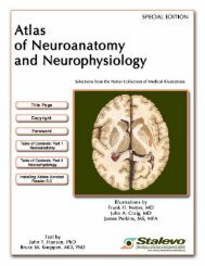
Fig. 2 – Immunohistochemistry for GFP and cell-specific markers in the acute phase of spinal cord injury. Near the lesion<br />
epicenter, GFP-positive round cells were also positive for neutrophil antigen (A–C, D, arrows). GFP-positive spindle-shaped cells<br />
negative for neutrophil antigen were observed (D, arrowheads). Some of the GFP-positive spindle-shaped cells were positive for<br />
vimentin, a marker for cells of the mesenchymal lineage (E, arrow). There was no significant difference in the number of<br />
GFP-positive cells between the SCI+PBS and SCI+G-<strong>CSF</strong> groups (F). The number of GFP- and neutrophil-double-positive cells<br />
was smaller in the SCI+G-<strong>CSF</strong> group than in the SCI+PBS group (F, hatched column, p