

PDF Version - Glidewell Dental Labs
PDF Version - Glidewell Dental Labs
PDF Version - Glidewell Dental Labs

Create successful ePaper yourself
Turn your PDF publications into a flip-book with our unique Google optimized e-Paper software.
3. Acrylic provisionals should be placed with<br />
Durelon (3M ESPE ; St. Paul, MN) temporary<br />
cement. Durelon is antimicrobial and helps<br />
decrease sensitivity.<br />
4. Remove provisional at time of surgery for<br />
access. Ideally, a mosquito forcep is used with<br />
a gentle rock at the incisal third of the occlusal<br />
surface of the provisional.<br />
5. Shape the tooth surface and remove old<br />
margin, as well as 360 degrees of CEJs. A<br />
flat-ended bur with a 4-degree taper is best for<br />
biologic shaping. A diamond grit is best.<br />
6. Correct any reverse architecture and remove<br />
any necessary bone where biologic width<br />
issues are still present. The goal is to create an<br />
osseous contour identical to the soft tissue<br />
contours that take place when forming a new<br />
biologic width.<br />
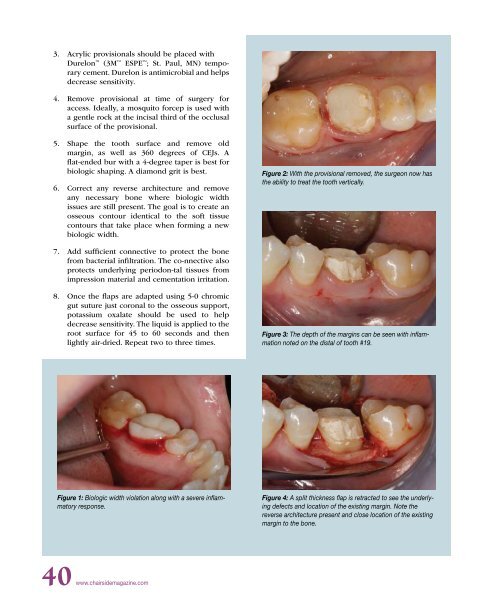
Figure 2: With the provisional removed, the surgeon now has<br />
the ability to treat the tooth vertically.<br />
7. Add sufficient connective to protect the bone<br />
from bacterial infiltration. The co-nnective also<br />
protects underlying periodon-tal tissues from<br />
impression material and cementation irritation.<br />
8. Once the flaps are adapted using 5-0 chromic<br />
gut suture just coronal to the osseous support,<br />
potassium oxalate should be used to help<br />
decrease sensitivity. The liquid is applied to the<br />
root surface for 45 to 60 seconds and then<br />
lightly air-dried. Repeat two to three times.<br />
Figure 3: The depth of the margins can be seen with inflammation<br />
noted on the distal of tooth #19.<br />
Figure 1: Biologic width violation along with a severe inflammatory<br />
response.<br />
Figure 4: A split thickness flap is retracted to see the underlying<br />
defects and location of the existing margin. Note the<br />
reverse architecture present and close location of the existing<br />
margin to the bone.<br />
40 www.chairsidemagazine.com


