PDF file: Annual Report 2002/2003 - Scottish Crop Research Institute
PDF file: Annual Report 2002/2003 - Scottish Crop Research Institute
PDF file: Annual Report 2002/2003 - Scottish Crop Research Institute

You also want an ePaper? Increase the reach of your titles
YUMPU automatically turns print PDFs into web optimized ePapers that Google loves.
Mechanisms & Processes<br />
Studying plasmodesmal targeting of TMV-MP<br />
using FRAP<br />
K.M. Wright, N.T. Wood 1 , K.MacKenzie 2 & K.J. Oparka<br />
Tobacco mosaic virus (TMV) is frequently studied<br />
as a model system to identify the mechanisms<br />
involved in plant viral infections. The structure and<br />
functions of all its genes have been identified and of<br />
particular interest is the 30-kDa protein required for<br />
cell-to-cell movement. This movement protein (MP)<br />
is involved in the transport of viral RNA to and<br />
through plasmodesmata (PD). TMV-MP accumulates<br />
within PDs and increases the size-exclusion limit of<br />
the PDs to allow trafficking of viral RNA to the next<br />
cell in the form of a MP-complex.<br />
In recent years it has been possible to produce TMV<br />
derivatives that express fully functional MP fused to<br />
green fluorescent protein (GFP) thus enabling visualisation<br />
of the MP. At the leading edge of an infection,<br />
and throughout the infection site, MP is located within<br />
PDs. Near the edge of an infection, MP is associated<br />
with vertices of the cortical endoplasmic reticulum<br />
(ER) whilst further in the MP can be seen aligning<br />
with the microtubules (MT). Some researchers have<br />
speculated that MT are involved in the targeting of<br />
TMV-MP to PDs. However, at SCRI we have shown<br />
that disruption of MT with pharmacological agents<br />
has no effect on lesion growth and we suggest that<br />
MT are instead involved in the degradation of MP<br />
later in the infection process.<br />
We are therefore investigating the targeting of MP to<br />
PDs using a technique called fluorescence recovery<br />
1<br />
after photobleaching (FRAP). Tobacco leaves are<br />
infected with TMV-MP-GFP virus and the edge of<br />
the infection site located using a confocal laser scanning<br />
microscope. Using the laser it is possible to<br />
bleach the fluorescent GFP attached to MP within a<br />
PD. Images are recorded and then measured as the<br />
fluorescence increases due to the movement of new<br />
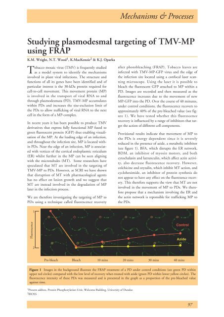
MP-GFP into the PD. Over the course of 40 minutes,<br />
under control conditions, the fluorescence recovers to<br />
approximately 40% of the pre-bleached value (see figure<br />
1). We have tested whether this fluorescence<br />
recovery is influenced by a range of inhibitors that target<br />
the action of different cell components.<br />
Provisional results indicate that movement of MP to<br />
the PDs is energy dependent since it is severely<br />
reduced in the presence of azide, a metabolic inhibitor<br />
(see figure 1). BFA, which disrupts the ER network,<br />
BDM, an inhibitor of myosin motors, and both<br />
cytochalasin and latrunculin, which affect actin activity,<br />
also decrease fluorescence recovery. However,<br />
colchicine and oryzalin, which inhibit MT action, and<br />
cycloheximide, an inhibitor of protein synthesis do<br />
not appear to have any effect on the fluoresence recovery.<br />
This therefore supports the view that MT are not<br />
involved in the movement of MP to PDs. We therefore<br />
propose that a mechanism involving the ER and<br />
the actin network is reponsible for trafficking MP to<br />
the PDs.<br />
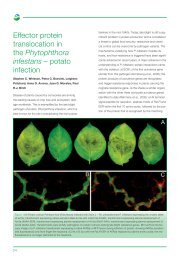
Fluorescence<br />
2<br />
Pre-bleach Bleach 10 mins 20 mins 30 mins 40 mins<br />
Figure 1 Images in the background illustrate the FRAP treatment of a PD under control conditions (see green PD within<br />
upper red circles) compared with the low level of recovery when treated with azide (green PD within lower yellow circles). The<br />
fluorescence intensity of these PDs was measured and is presented in the graph as a proportion of the pre-bleached value<br />
against time.<br />
1 Present address, Protein Phosphorylation Unit, Welcome Building, University of Dundee<br />
2 BIOSS<br />
97