SummEr/FAll 2011 - Nazareth College
SummEr/FAll 2011 - Nazareth College
SummEr/FAll 2011 - Nazareth College

Create successful ePaper yourself
Turn your PDF publications into a flip-book with our unique Google optimized e-Paper software.
LIFE | of the mind<br />
Biology Student Research<br />
Reaches New Dimension<br />
by Matthew Temple<br />
“S<br />
eeing is believing” has now taken on new<br />
levels of meaning in the biology department<br />
at <strong>Nazareth</strong> with a new microscope<br />
that “sees” in 3-D. I’ve been teaching<br />
<strong>Nazareth</strong> students how to look at cells<br />
through a microscope since 1984. And since<br />
1984, I’ve often had to apologize to them<br />
because we know that cells actually have<br />
three-dimensional shapes—as spheres or cubes<br />
or even blobs—but under the microscope,<br />
those cells are often flattened in order to be<br />
examined. It’s like the difference between<br />
looking at a fully inflated soccer ball and one<br />
that has been deflated into a flat and distorted<br />
caricature of its former self. But at <strong>Nazareth</strong>,<br />
this is now no longer the case.<br />
Last summer, the biology department<br />
acquired a microscope that can “see” into a<br />
cell in three dimensions and that enables us to<br />
appreciate how cells package vital components<br />
within their spaces. Technically, this is called an<br />
optical-sectioning microscope, because it takes<br />
finely focused pictures of up to 50 slices of a<br />
cell from top to bottom. Those slices are then<br />
compiled by a computer into a three-dimensional<br />
rendering of that cell. Furthermore, different<br />
components within a cell are literally lit<br />
up by fluorescent dyes, which can make DNA<br />
(the genetic material) glow a brilliant blue, fat<br />
droplets (cells have to deal with fat, too) a vivid<br />
green, and structural fibers a deep red.<br />
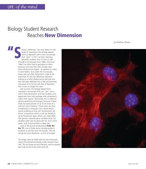
Look at the picture produced by Kelly Noble<br />
’11, one of three senior biology research<br />
students to use the new microscope. The red<br />
strings are actin filaments—a kind of bungee<br />
This image, taken by Noble with the new microscope,<br />
shows three-dimensional details inside cultured mouse<br />
cells. The red strings are actin filaments, and the spotted<br />
blue ovals are the nuclei within each cell.<br />
28 CONNECTIONS | Summer/Fall <strong>2011</strong> www.naz.edu