SummEr/FAll 2011 - Nazareth College
SummEr/FAll 2011 - Nazareth College
SummEr/FAll 2011 - Nazareth College

You also want an ePaper? Increase the reach of your titles
YUMPU automatically turns print PDFs into web optimized ePapers that Google loves.
“…few students in other undergraduate biology departments<br />
have such free access to such a powerful instrument.”<br />
cord—inside cultured mouse cells. The blue ovals with bright spots are<br />
the DNA-rich nuclei within each cell. Kelly’s work suggests that actin<br />
has a unique shape around and within the nucleus. Shaughna Szymanski<br />
’11 used this microscope to explore how mouse cells store excess<br />
fat, while Jessica Reeves ’11 showed that cells develop unusual<br />
nuclear shapes in response to a potent chemical. Their work would not<br />
have been possible at <strong>Nazareth</strong> even a year ago. Better yet, their work<br />
provides a solid foundation for students and faculty at <strong>Nazareth</strong> to use<br />
this new microscope to its fullest capacity in laboratory classes and in<br />
collaborative research.<br />
The idea for getting this kind of microscope at <strong>Nazareth</strong> started<br />
years ago, on a sabbatical. One of many great things about teaching<br />
at <strong>Nazareth</strong> is the opportunity for a sabbatical every seven years. So<br />
far, I’ve had three sabbaticals: each has involved at least a semester<br />
of research at the Jackson Laboratory, a world-class genetics research<br />
institute in Bar Harbor, Maine. For a geneticist who wants to learn the<br />
latest techniques and work with some of the greatest researchers, the<br />
Jackson Lab is the place to go. My last two sabbaticals gave me the<br />
opportunity to use those microscopes that can see inside cells in 3-D.<br />
This technology was literally an eye-opener for me: I could see inside<br />
the nuclei of cells (where genes are) in a radically different way. At<br />
“The JAX,” as it is called, I watched chromosomes in trouble in mutant<br />
mice and looked at a protein that seemed to hold certain genes close<br />
to the wall of the cell’s nucleus.<br />
Using these sophisticated microscopes made me wonder how my<br />
students at <strong>Nazareth</strong> could see cells this way as well. Early in 2010,<br />
Deborah Dooley ’75, dean of the <strong>College</strong> of Arts and Sciences, asked<br />
science faculty for suggestions for equipment for the new Integrated<br />
Center for Math and Science—then only on the horizon but now under<br />
construction. I suggested an optical-sectioning microscope much<br />
like the one I had used at the Jackson Lab. Dean Dooley found the<br />
funds for it, and <strong>Nazareth</strong>’s Information Technology Services provided<br />
the computer that runs the microscope and produces its 3-D images.<br />
By August of last year, the new microscope and its computer were<br />
ready for our cell biology and senior research students. And by April<br />
<strong>2011</strong>, Kelly, Shaunghna, and Jessica had used it to describe the shapes<br />
of DNA, fats, and protein filaments inside cells in brilliant color and<br />
accurate detail.<br />
As far as I can tell, few students in other undergraduate biology<br />
departments have such free access to such a powerful instrument.<br />
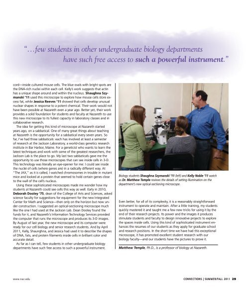
Biology students Shaughna Szymanski ’11 (left) and Kelly Noble ’11 watch<br />
as Dr. Matthew Temple reviews the details of setting illumination on the<br />
department’s new optical-sectioning microscope.<br />
Even better, for all of its complexity, it is a reasonably straightforward<br />
instrument to operate and maintain. After a little training, my students<br />
quickly mastered it and taught me a few new tricks for using it by the<br />
end of their research projects. Its power and the images it produces<br />
stimulate students and faculty to design innovative projects to explore<br />
the spaces inside cells. Using this kind of sophisticated instrument enhances<br />
the resumes of our students as they apply for graduate school<br />
and research positions. In the short time we have had this exceptional<br />
microscope, it has promoted excellent student research with our<br />
biology faculty—and our students have the pictures to prove it.<br />
Matthew Temple, Ph.D., is a professor of biology at <strong>Nazareth</strong>.<br />
www.naz.edu CONNECTIONS | Summer/Fall <strong>2011</strong> 29