Miniaturized Ultrasound Imaging Probes Enabled by CMUT Arrays ...
Miniaturized Ultrasound Imaging Probes Enabled by CMUT Arrays ...
Miniaturized Ultrasound Imaging Probes Enabled by CMUT Arrays ...

Create successful ePaper yourself
Turn your PDF publications into a flip-book with our unique Google optimized e-Paper software.
32nd Annual International Conference of the IEEE EMBS<br />
Buenos Aires, Argentina, August 31 - September 4, 2010<br />
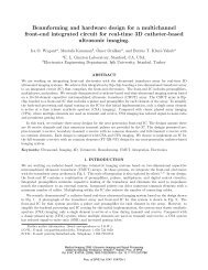
<strong>Miniaturized</strong> <strong>Ultrasound</strong> <strong>Imaging</strong> <strong>Probes</strong> <strong>Enabled</strong> <strong>by</strong> <strong>CMUT</strong><br />
<strong>Arrays</strong> with Integrated Frontend Electronic Circuits<br />
B. (Pierre) T. Khuri-Yakub, Ömer Oralkan, Amin Nikoozadeh, Ira O. Wygant, Steve Zhuang, Mustafa<br />
Gencel, Jung Woo Choe, Douglas N. Stephens, Alan de la Rama, Peter Chen, Feng Lin, Aaron<br />
Dentinger, Douglas Wildes, Kai Thomenius, Kalyanam Shivkumar, Aman Mahajan, Chi Hyung Seo,<br />
Matthew O’Donnell, Uyen Truong, and David J. Sahn<br />
Abstract— Capacitive micromachined ultrasonic transducer<br />
(<strong>CMUT</strong>) arrays are conveniently integrated with frontend<br />
integrated circuits either monolithically or in a hybrid<br />
multichip form. This integration helps with reducing the<br />
number of active data processing channels for 2D arrays. This<br />
approach also preserves the signal integrity for arrays with<br />
small elements. Therefore <strong>CMUT</strong> arrays integrated with<br />
electronic circuits are most suitable to implement miniaturized<br />
probes required for many intravascular, intracardiac, and<br />
endoscopic applications. This paper presents examples of<br />
miniaturized <strong>CMUT</strong> probes utilizing 1D, 2D, and ring arrays<br />
with integrated electronics.<br />
I. INTRODUCTION<br />
In conventional ultrasound imaging systems that utilize<br />
1D transducer arrays for 2D cross-sectional imaging the<br />
frontend transmit/receive (T/R) electronic circuits are<br />
typically located in the main processing unit. The transducer<br />
array is packaged in the form of a hand-held probe. Each<br />
element in the array is connected to a dedicated frontend<br />
channel through a long (~2 m) coaxial cable.<br />
This conventional scheme has not been practical for<br />
transducer arrays with increased number of elements such as<br />
the 2D arrays with over 2000 elements. All major ultrasound<br />
system providers today offer 2D matrix arrays for 3D<br />
imaging. All these 2D matrix probes house a portion of the<br />
beamforming electronics next to the array so that the number<br />
of cables between the system and the transducer can be<br />
minimized and the acquired data can be processed <strong>by</strong> the<br />
existing imaging systems without having the need for<br />
thousands of active electronic channels. Placing the frontend<br />
electronics next to the array also improves the signal<br />
This work was supported <strong>by</strong> the National Institutes of Health under<br />
grants CA99059, CA134720, and HL67647.<br />
Ö.O., A.N., I.O.W., S.Z., M.G., J-W.C., and P.T.K-Y. are with the E. L.<br />
Ginzton Laboratory, Stanford University, Stanford, CA 94305, USA (email:<br />
ooralkan@stanford.edu).<br />
D.N.S. is with the University of California, Davis, CA 95616, USA.<br />
A.D.L.R. and P.C. are with St. Jude Medical, Irvine, CA 92618, USA.<br />
F.L., A.D., D.W., and K.T. are with the General Electric Global<br />
Research Center, Niskayuna, NY 12309, USA.<br />
K.S. and A.M. are with the University of California, Los Angeles, CA<br />
90095, USA.<br />
C.H.S. and M.O. are with the University of Washington, Seattle, WA<br />
98101, USA.<br />
U.T. and D.J.S. are with the Oregon Health and Science University,<br />
Portland, OR 97239, USA.<br />
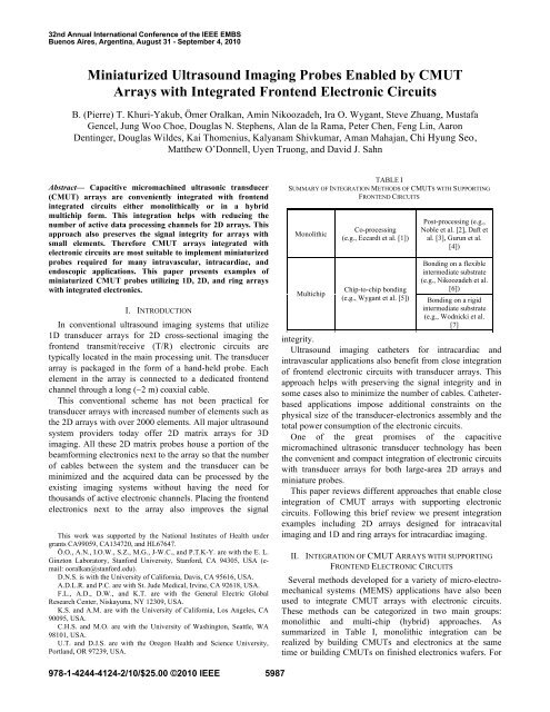
TABLE I<br />
SUMMARY OF INTEGRATION METHODS OF <strong>CMUT</strong>S WITH SUPPORTING<br />
FRONTEND CIRCUITS<br />
Monolithic<br />
Multichip<br />
Co-processing<br />
(e.g., Eccardt et al. [1])<br />
Chip-to-chip bonding<br />
(e.g., Wygant et al. [5])<br />
Post-processing (e.g.,<br />
Noble et al. [2], Daft et<br />
al. [3], Gurun et al.<br />
[4])<br />
Bonding on a flexible<br />
intermediate substrate<br />
(e.g., Nikoozadeh et al.<br />
[6])<br />
Bonding on a rigid<br />
intermediate substrate<br />
(e.g., Wodnicki et al.<br />
[7]<br />
integrity.<br />
<strong>Ultrasound</strong> imaging catheters for intracardiac and<br />
intravascular applications also benefit from close integration<br />
of frontend electronic circuits with transducer arrays. This<br />
approach helps with preserving the signal integrity and in<br />
some cases also to minimize the number of cables. Catheterbased<br />
applications impose additional constraints on the<br />
physical size of the transducer-electronics assembly and the<br />
total power consumption of the electronic circuits.<br />
One of the great promises of the capacitive<br />
micromachined ultrasonic transducer technology has been<br />
the convenient and compact integration of electronic circuits<br />
with transducer arrays for both large-area 2D arrays and<br />
miniature probes.<br />
This paper reviews different approaches that enable close<br />
integration of <strong>CMUT</strong> arrays with supporting electronic<br />
circuits. Following this brief review we present integration<br />
examples including 2D arrays designed for intracavital<br />
imaging and 1D and ring arrays for intracardiac imaging.<br />
II. INTEGRATION OF <strong>CMUT</strong> ARRAYS WITH SUPPORTING<br />
FRONTEND ELECTRONIC CIRCUITS<br />
Several methods developed for a variety of micro-electromechanical<br />
systems (MEMS) applications have also been<br />
used to integrate <strong>CMUT</strong> arrays with electronic circuits.<br />
These methods can be categorized in two main groups:<br />
monolithic and multi-chip (hybrid) approaches. As<br />
summarized in Table I, monolithic integration can be<br />
realized <strong>by</strong> building <strong>CMUT</strong>s and electronics at the same<br />
time or building <strong>CMUT</strong>s on finished electronics wafers. For<br />
978-1-4244-4124-2/10/$25.00 ©2010 IEEE 5987
multichip (hybrid) integration an intermediate substrate may<br />
or may not be used.<br />
A. Co-Processing<br />
A BiCMOS process using 16 masks has been used with<br />
only minor modifications including an additional<br />
photolithography step and sacrificial layer etching to<br />
fabricate <strong>CMUT</strong>s side-<strong>by</strong>-side with electronic circuits on the<br />
same substrate [1]. Although this method offers a costeffective<br />
means of integration, it has two major limitations:<br />
1) The transducer element area is shared <strong>by</strong> electronic<br />
circuits or interconnects. 2) The device dimensions in the<br />
vertical direction are limited <strong>by</strong> the film thicknesses<br />
available in the process used to make electronic circuits.<br />
dedicated T/R frontend circuitry, only one channel was<br />
programmed to be active at a time for simplicity and hence<br />
classical synthetic aperture imaging was performed with this<br />
first-generation prototype.<br />
B. Post-Processing<br />
Another monolithic integration technique is to make the<br />
electronic circuits first using a foundry process and then<br />
build <strong>CMUT</strong>s on top of finished electronics <strong>by</strong> further<br />
processing, which includes surface passivation,<br />
planarization, opening of contacts to electronics, and further<br />
successive thin film deposition and etching steps to define<br />
the <strong>CMUT</strong> membranes [2, 3, 4]. This approach has good<br />
area utilization. However, processing techniques for making<br />
<strong>CMUT</strong>s are still limited, mainly due to temperature<br />
constraints set <strong>by</strong> the existing metal lines on the electronics.<br />
C. Independently Processing<br />
By making <strong>CMUT</strong>s and electronic circuits on two<br />
separate substrates one can use optimized process flows for<br />
each component. <strong>CMUT</strong>s, in this case, need through-wafer<br />
via interconnections from the front side of the substrate to<br />
the back side. Deep reactive ion etching (DRIE) enables<br />
these vias in silicon. <strong>CMUT</strong> arrays can then be bonded<br />
directly on electronics [5] or on an intermediate substrate,<br />
which can be flexible [6] or rigid [7]. Direct bonding usually<br />
requires that the electronics die area to be greater than the<br />
<strong>CMUT</strong> die area. Using an intermediate substrate makes the<br />
size of the <strong>CMUT</strong> and electronics dice independent. Both<br />
components can be tiled to obtain larger arrays. Flexible<br />
substrates are preferred for catheter applications because of<br />
the smaller form factor they offer through bending.<br />
III. 2D <strong>CMUT</strong> ARRAYS WITH INTEGRATED FRONTEND<br />
ELECTRONIC CIRCUITS<br />
We developed two generations of frontend integrated<br />
circuits for 16x16-element 2D <strong>CMUT</strong> arrays for 3D<br />
intracavital imaging applications such as endoscopic,<br />
endorectal, or endovaginal imaging.<br />
A. Synthetic Aperture System<br />
The first generation frontend integrated circuit (IC) we<br />
designed comprised a 25-V pulser, a T/R switch and a lownoise<br />
preamplifier for each transducer element in the array<br />
[Fig.1(a)] [5]. Both the 2D <strong>CMUT</strong> array and the frontend IC<br />
had a pitch of 250 µm. Using Sn-Pb eutectic solder bumps<br />
the two components were directly bonded together<br />
[Fig.1(b)]. Although each transducer element had their<br />
Fig. 1 (a) Custom-designed frontend IC for the 3D synthetic<br />
aperture system. (b) A 16x16-element, 250-µm pitch, 2D<br />
<strong>CMUT</strong> array bonded on frontend electronics.<br />
B. Full-Transmit X-Receive System with Integrated<br />
Beamforming Hardware<br />
The second-generation frontend IC we designed for the<br />
16x16-element 2D array included digital circuitry to perform<br />
the functions of a transmit beamformer on the chip [Fig.2(a)]<br />
[8]. With this IC 224 elements can be simultaneously fired in<br />
transmit and 16 elements can be used at a time for receive.<br />
We use a beamforming scheme, which we developed to<br />
reduce the frontend hardware complexity with minimum<br />
compromise from image quality. In this scheme the 32<br />
diagonal elements in the array constitute the receive aperture<br />
and the remainder of the array constitutes the transmit<br />
aperture. A preamplifier is provided for each of the 32<br />
receive elements. To interface to a 16-channel data<br />
acquisition system, 16 of the 32 receive elements are active<br />
at a time. Sixteen buffers drive the cable capacitance in<br />
receive. A high-voltage pulser and an 8-bit shift register are<br />
provided for each of the 224 transmitting elements. The<br />
timing of each pulser is determined <strong>by</strong> delay information<br />
stored in the shift register. When transmitting, a particular<br />
pulser fires when a global counter equals the pulser’s stored<br />
delay value. For each new beam, delay information is loaded<br />
into the delay shift registers <strong>by</strong> 16 parallel lines. This<br />
scheme also reduces the number of cables between the<br />
backend system and the frontend. We bonded a 16x16-<br />
5988
element 2D <strong>CMUT</strong> array operating at 2.5 MHz directly on<br />
this frontend IC and used this prototype for imaging several<br />
phantoms in 3D (Fig.3).<br />
IV. INTRACARDIAC ECHOGRAPHY CATHETERS WITH <strong>CMUT</strong><br />
ARRAYS WITH INTEGRATED FRONTEND ELECTRONIC<br />
CIRCUITS<br />
We developed two types of forward looking intracardiac<br />
echography (ICE) catheters to provide guidance during<br />
electrophysiological (EP) interventions.<br />
A. Forward-Looking Microlinear <strong>Arrays</strong> for Real-Time<br />
Cross-Sectional <strong>Imaging</strong><br />
The first device we developed for forward-looking ICE<br />
applications is a 24-element microlinear array operating at<br />
10 MHz center frequency with a fractional bandwidth of<br />
100%. The element pitch is 63 µm. The array measures 1.7<br />
mm <strong>by</strong> 1.3 mm and employs doped-polysilicon filled<br />
through-wafer via interconnects [Fig.4(a)]. This array was<br />
bonded on one side of a custom flexible printed circuit board<br />
(PCB). The 24-channel electronics die providing each<br />
transducer element with a T/R switch, a low-noise<br />
preamplifier and an output buffer was bonded on the back<br />
side of the flexible PCB [Fig.4(b)]. With this IC the transmit<br />
pulses with amplitudes up to -/+ 50 V were provided from<br />
the imaging system (GE Vivid7), which is connected to the<br />
flex assembly <strong>by</strong> microcoaxial cables. A thin layer of<br />
Polydimethylsiloxane (PDMS) provided electrical insulation<br />
on the front side of the array.<br />
The final catheter packaged in a 9-F shaft [Fig.4(c)] was<br />
tested successfully in an animal study recently providing<br />
forward-looking cross-sectional images up to 4-cm depth<br />
(Fig.5).<br />
Fig.2. (a) Custom-designed frontend IC for the full-transmit X-<br />
receive imaging system. (b) A 16x16-element, 250-µm pitch,<br />
2D <strong>CMUT</strong> array bonded on frontend electronics.<br />
Fig. 4. (a) 24-element <strong>CMUT</strong> microlinear array. (b) The flex<br />
assembly with <strong>CMUT</strong> microlinear array bonded on side and<br />
the frontend IC on the other. (c) The final 9F catheter with<br />
forward-looking microlinear <strong>CMUT</strong> array.<br />
Fig .3. 3D renedered image of a nylon wire phantom obtained<br />
with a 2.5-MHz 2D <strong>CMUT</strong> array<br />
Fig. 5. A 2D image obtained with the <strong>CMUT</strong> ML array in-vivo<br />
in a pig heart.<br />
5989
B. Forward-Looking Ring <strong>Arrays</strong> for Volumetric <strong>Imaging</strong><br />
The second type of the ICE catheters we developed is<br />
based on a 10-MHz, 64-element <strong>CMUT</strong> ring array<br />
[Fig.6(a)]. The electronics we used along with this array has<br />
the same circuit topology as the one used with the<br />
microlinear array. However, in this case 8 channels are<br />
grouped on one die and 8 ICs were used to address the entire<br />
array. The <strong>CMUT</strong> ring array and the 8 ICs were bonded on<br />
an 8-legged flexible printed circuit board. A compact<br />
assembly was achieved <strong>by</strong> folding the legs of the flex circuit<br />
on a support structure [Fig.6(b)]. Microcoaxial cables<br />
connected this assembly to the imaging system. A<br />
programmable imaging platform (Verasonics, Redmond,<br />
WA) was used to reconstruct real-time volume images on<br />
multi-plane display with the option of off-line 3D rendering.<br />
The initial prototype was packaged in a 12-F catheter shaft<br />
[Fig.6(c)] and was used for imaging several phantoms (Fig.<br />
7).<br />
The ring catheter provides a 4.5-F inner lumen that can be<br />
used to introduce therapeutic devices such as RF ablation<br />
catheters. Optical fibers can be inserted into the inner lumen<br />
to enable photoacoustic imaging.<br />
V. CONCLUSION<br />
We have demonstrated several prototypes of miniaturized<br />
ultrasound probes using <strong>CMUT</strong> arrays with integrated<br />
electronics. The use of standard silicon microfabrication<br />
techniques in <strong>CMUT</strong> manufacturing enables densely<br />
populated transducer arrays with arbitrary geometries. These<br />
arrays can be conveniently integrated with supporting<br />
Fig. 6. (a) 64-element <strong>CMUT</strong> ring array. (b) Flex assembly<br />
with the <strong>CMUT</strong> array and eight 8-channel frontend ICs. (c)<br />
The final 12-F catheter.<br />
Fig. 7. 3D rendered image of a metal spring obtained with the<br />
<strong>CMUT</strong> ring array.<br />
electronic circuits monolithically or in the form of a<br />
multichip module to reduce the number of cables between<br />
the probe and the system and also to preserve signal<br />
integrity. Results presented in this paper show that <strong>CMUT</strong><br />
technology has the potential to enable many applications in<br />
ultrasound imaging and therapy that were not possible with<br />
the conventional piezoelectric transducers.<br />
ACKNOWLEDGMENT<br />
Authors thank National Semiconductor for manufacturing<br />
the frontend integrated circuits, Tyco Electronics for cabling,<br />
and PacTech USA for their support with packaging.<br />
REFERENCES<br />
[1] P. C. Eccardt, K. Niederer, and B. Fischer , “Micromachined<br />
transducers for ultrasound applications," Proc. IEEE Ultrason. Symp.,<br />
pp. 1609-1618, 1997.<br />
[2] R. A. Noble, R. R. Davies, D. O. King, M. M. Day, A. R. D. Jones, J.<br />
S. McIntosh, D. A. Hutchins, and P. Saul, “Low temperature<br />
micromachined cMUTs with fully-integrated analogue front-end<br />
electronics,” Proc. IEEE Ultrason. Symp., pp. 1045–1050, 2002.<br />
[3] C. Daft, S. Calmes, D. da Graca, K. Patel, P. Wagner, and I.<br />
Ladabaum, “Microfabricated ultrasonic transducers monolithically<br />
integrated with high voltage electronics,” Proc. IEEE Ultrason.<br />
Symp., pp. 493–496, 2004.<br />
[4] G. Gurun, M. S. Qureshi, M. Balantekin, R. Guldiken, J. Zahorian, P.<br />
Sheng-Yu, A. Basu, M. Karaman, P. Hasler, L. Degertekin, “Frontend<br />
CMOS electronics for monolithic integration with <strong>CMUT</strong> arrays:<br />
circuit design and initial experimental results,” Proc. IEEE Ultrason.<br />
Symp., pp. 390-393, 2008.<br />
[5] I. Wygant, X. Zhuang, D. Yeh, Ö. Oralkan, A. S. Ergun, M. Karaman,<br />
and B. T. Khuri-Yakub, “Integration of 2D <strong>CMUT</strong> arrays with frontend<br />
electronics for volumetric ultrasound imaging,” IEEE Trans.<br />
Ultrason., Ferroelect., Freq. Contr., vol. 55, no. 2, pp. 327-342, Feb.<br />
2008.<br />
[6] A. Nikoozadeh, Ö. Oralkan, M. Gencel, J. W. Choe, D. N. Stephens,<br />
A. de la Rama, P. Chen, K. Thomenius, A. Dentinger, D. Wildes, K.<br />
Shivkumar, A. Mahajan, M. O’Donnell, D. Sahn and P. T. Khuri-<br />
Yakub, “Forward-Looking Volumetric Intracardiac <strong>Imaging</strong> Using a<br />
Fully Integrated <strong>CMUT</strong> Ring Array,” presented at the IEEE Ultrason.<br />
Symp., Rome, Italy, Sep. 2009.<br />
[7] R. Wodnicki, C. G. Woychik, A. T. Byun, R. Fisher, K. Thomenius,<br />
D.-S. Lin, X. Zhuang, O. Oralkan, S. Vaithilingam and B. T. Khuri-<br />
Yakub, “Multi-row linear cMUT array using cMUTs and multiplexing<br />
electronics,” presented at the IEEE Ultrason. Symp., Rome, Italy, Sep.<br />
2009.<br />
[8] I. O. Wygant, N. S. Jamal, H. J. Lee, A. Nikoozadeh, Ö. Oralkan, M.<br />
Karaman, and B. T. Khuri-Yakub, “An integrated circuit with transmit<br />
beamforming flip-chip bonded to a 2-D <strong>CMUT</strong> array for 3-D<br />
ultrasound imaging,” IEEE Trans. Ultrason., Ferroelect., Freq.<br />
Contr., vol. 56, no. 10, pp. 2145-2156, Oct. 2009.<br />
5990