Undergraduate Research Journal
Undergraduate Research Journal
Undergraduate Research Journal
You also want an ePaper? Increase the reach of your titles
YUMPU automatically turns print PDFs into web optimized ePapers that Google loves.
Molecular Analysis of the Interaction Between Staphylococcus aureus Protein Sbi and Immune System Protein C3d<br />
Wilson Rodriguez<br />
collection of independent and communicating scripts and<br />
is written in the R software environment for statistical<br />
computing. AESOP performs alanine scans, calculates<br />
electrostatic potentials using the Poisson-Boltzmann<br />
equation, determines electrostatic similarity distances<br />
(ESD), performs hierarchical electrostatic clustering of<br />
ESDs, calculates electrostatic free energies of association,<br />
and provides a graphical representation of electrostatic<br />
potentials. AESOP’s centralized functions have facilitated<br />
the study of electrostatics in the function of protein-protein<br />
interactions. 5-9<br />
The AESOP protocol begins with acquiring a file with<br />
the coordinates of the crystal structure from the Protein<br />
Data Bank (PDB), which is visually inspected for missing<br />
or duplicate residues. Duplicates were removed and no<br />
missing residues were found. It is then cleaned by removing<br />
the header and footer of the file and any heteroatoms<br />
and water atoms the structure may have. Once the PDB<br />
file is processed, AESOP performs the aforementioned<br />
automated tasks. It starts by reading in the coordinates of<br />
the PDB file and changes its format into a PQR file, using<br />
the PDB2PQR program. 1,2,4 PQR files are very similar<br />
to PDB files except that in addition to coordinates, they<br />
contain atomic charges and radii. If hydrogen atoms are<br />
missing in PDB files, which are typical of crystallographic<br />
structures, PDB2PQR also adds hydrogen atoms. The next<br />
step is to generate a family of mutants from the parent<br />
PQR file. AESOP does so by reading the protein sequence<br />
residues one at a time in order to locate one of the five<br />
charged residues of interest, histidines, arginines, lysines,<br />
aspartates or glutamates, and then truncates the side chain<br />
to the beta carbon, shortens the gamma carbon’s bond and<br />
changes it into a hydrogen with correct bond length and<br />
atomic charge. Once the mutation is made, a new PQR<br />
file is saved and stored for further calculations. The script<br />
then returns to the parent PQR to search and locate the<br />
next available charged residue to mutate to alanine until<br />
it has checked and mutated all the residues in the parent<br />
sequence. This process is called computational alanine<br />
scan and each of the mutant proteins has a single residue<br />
mutation generated from the parent protein. In the C3d-<br />
Sbi complex there were 94 total residue mutations, 64<br />
belonging to C3d and 30 belonging to Sbi.<br />
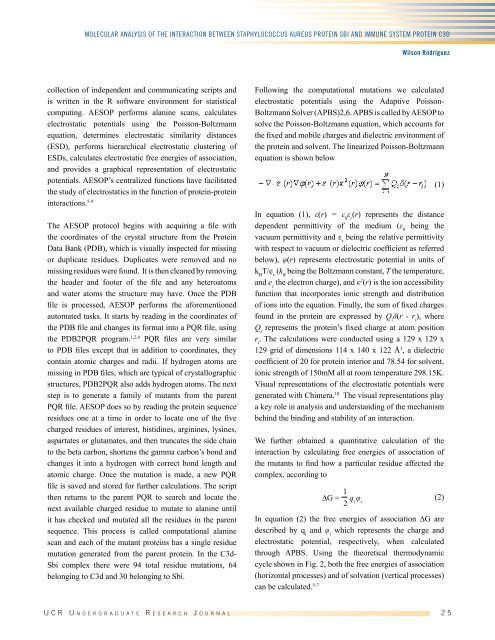
Following the computational mutations we calculated<br />
electrostatic potentials using the Adaptive Poisson-<br />
Boltzmann Solver (APBS)2,6. APBS is called by AESOP to<br />
solve the Poisson-Boltzmann equation, which accounts for<br />
the fixed and mobile charges and dielectric environment of<br />
the protein and solvent. The linearized Poisson-Boltzmann<br />
equation is shown below<br />
In equation (1), ε(r) = ε 0<br />
ε r<br />
(r) represents the distance<br />
dependent permittivity of the medium (ε 0<br />
being the<br />
vacuum permittivity and ε r<br />
being the relative permittivity<br />
with respect to vacuum or dielectric coefficient as referred<br />
below), φ(r) represents electrostatic potential in units of<br />
k B<br />
T/e c<br />
(k B<br />
being the Boltzmann constant, T the temperature,<br />
and e c<br />
the electron charge), and к 2 (r) is the ion accessibility<br />
function that incorporates ionic strength and distribution<br />
of ions into the equation. Finally, the sum of fixed charges<br />
found in the protein are expressed by Q i<br />
δ(r - r i<br />
), where<br />
Q i<br />
represents the protein’s fixed charge at atom position<br />
r i<br />
. The calculations were conducted using a 129 x 129 x<br />
129 grid of dimensions 114 x 140 x 122 Å 3 , a dielectric<br />
coefficient of 20 for protein interior and 78.54 for solvent,<br />
ionic strength of 150mM all at room temperature 298.15K.<br />
Visual representations of the electrostatic potentials were<br />
generated with Chimera. 10 The visual representations play<br />
a key role in analysis and understanding of the mechanism<br />
behind the binding and stability of an interaction.<br />
We further obtained a quantitative calculation of the<br />
interaction by calculating free energies of association of<br />
the mutants to find how a particular residue affected the<br />
complex, according to<br />
(1)<br />
∆G = 1<br />
2 q i φ i<br />
(2)<br />
In equation (2) the free energies of association ∆G are<br />
described by q i<br />
and φ i<br />
which represents the charge and<br />
electrostatic potential, respectively, when calculated<br />
through APBS. Using the theoretical thermodynamic<br />
cycle shown in Fig. 2, both the free energies of association<br />
(horizontal processes) and of solvation (vertical processes)<br />
can be calculated. 5-7<br />
U C R U n d e r g r a d u a t e R e s e a r c h J o u r n a l 2 5


