in vivo
in vivo
in vivo

Create successful ePaper yourself
Turn your PDF publications into a flip-book with our unique Google optimized e-Paper software.
Laboratory for<br />
Lymphocyte Development<br />
Thymic microenvironments critically support<br />
the development of T lymphocytes.<br />
The stromal cell types compos<strong>in</strong>g these<br />
microenvironments are epithelial <strong>in</strong> orig<strong>in</strong>, and<br />
form as non-polarized cells a three-dimensional<br />
(3-D) organized network (Fig.1).<br />
This type of organization is unique for<br />
epithelial cells <strong>in</strong> the thymus, because <strong>in</strong> other<br />
organs epithelial cells are polarized and placed<br />
on a basal lam<strong>in</strong>a form<strong>in</strong>g sheets of cells. The<br />
3-D organization of thymic epithelial cells (TECs)<br />
forms the basis of microenvironments, allow<strong>in</strong>g<br />
both for migration of thymocytes through<br />
the epithelial network, as well as for lymphostromal<br />
<strong>in</strong>teraction. In this way, TECs control<br />
various steps <strong>in</strong> T cell development, like clonal<br />
expansion, and also positive and negative<br />
selection. Reciprocally, the <strong>in</strong>tegrity of thymic<br />
microenvironments depends on the physical<br />
presence of thymocytes with<strong>in</strong> the epithelial<br />
network. We have previously shown that removal<br />
of thymocytes from the thymic environment,<br />
either by genetic manipulation or by biochemical<br />
methods leads to a dramatic shift <strong>in</strong> the<br />
phenotype and organization of TECs, <strong>in</strong>duc<strong>in</strong>g<br />
the formation of epithelial cell types which are<br />
normally found <strong>in</strong> the gastro-<strong>in</strong>test<strong>in</strong>al tract and<br />
the respiratory tract (Fig.2). These experiments<br />
have uncovered a mutual <strong>in</strong>terdependency<br />
between TECs and thymocytes, a phenomenon<br />
designated as “thymic crosstalk” (Fig.3). Thus,<br />
thymic crosstalk regulates the <strong>in</strong>tegrity and<br />
ma<strong>in</strong>tenance of the thymic stroma, and <strong>in</strong> this<br />
respect, thymocytes cont<strong>in</strong>uously promote their<br />
own development (see references 1-4).<br />
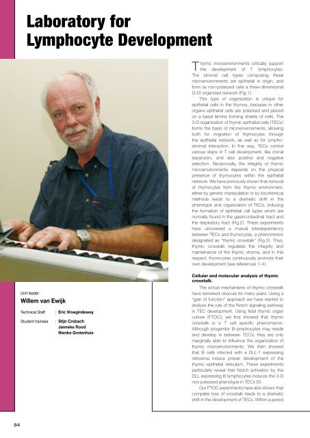
Unit leader<br />
Willem van Ewijk<br />
Technical Staff<br />
Student tra<strong>in</strong>ees<br />
: Eric Vroeg<strong>in</strong>dewey<br />
: Stijn Crobach<br />
Janneke Rood<br />
Nienke Grotenhuis<br />
Cellular and molecular analysis of thymic<br />
crosstalk.<br />
The actual mechanisms of thymic crosstalk<br />
have rema<strong>in</strong>ed obscure for many years. Us<strong>in</strong>g a<br />
“ga<strong>in</strong> of function” approach we have started to<br />
analyze the role of the Notch signal<strong>in</strong>g pathway<br />
<strong>in</strong> TEC development. Us<strong>in</strong>g fetal thymic organ<br />
culture (FTOC), we first showed that thymic<br />
crosstalk is a T cell specific phenomenon.<br />
Although progenitor B-lymphocytes may reside<br />
and develop <strong>in</strong> between TEC’s, they are only<br />
marg<strong>in</strong>ally able to <strong>in</strong>fluence the organization of<br />
thymic microenvironments. We then showed<br />
that B cells <strong>in</strong>fected with a DLL-1 express<strong>in</strong>g<br />
retrovirus <strong>in</strong>duce proper development of the<br />
thymic epithelial reticulum. These experiments<br />
particularly reveal that Notch activation by the<br />
DLL express<strong>in</strong>g B lymphocytes <strong>in</strong>duces the 3-D<br />
non polarized phenotype <strong>in</strong> TECs (5).<br />
Our FTOC experiments have also shown that<br />
complete loss of crosstalk leads to a dramatic<br />
shift <strong>in</strong> the development of TECs. With<strong>in</strong> a period<br />
84