Molluscan Research: Techniques for collecting, handling, preparing ...
Molluscan Research: Techniques for collecting, handling, preparing ...
Molluscan Research: Techniques for collecting, handling, preparing ...

You also want an ePaper? Increase the reach of your titles
YUMPU automatically turns print PDFs into web optimized ePapers that Google loves.
22<br />
determination of the position of the shell, facilitating<br />
comparison. Protoconchs should be shown in apical view, at<br />
right angles to the coiling axis or parallel to the coiling axis<br />
with the transition proto- to teleoconch in the centre.<br />
Bivalves should be shown with the outline of the shell<br />
parallel to the image plane and the prodissoconch (with at<br />
least long axis parallel to image plane) should be shown in<br />
umbonal view.<br />
SEM preparation of animals<br />
External anatomy can provide many useful features. The<br />
animal should be preserved in as natural a state as possible,<br />
ideally following relaxation. In general, <strong>for</strong>malin or<br />
glutaraldehyde fixed animals dry better than those fixed in<br />
ethanol only. Additional post-fixation may be carried out<br />
with osmium tetroxide (OsO 4 ), particularly if specimens are<br />
viewed in older SEMs that necessitate high accelerating<br />
voltages as OsO 4 will add conductivity to the specimen (see<br />
Appendix 1). Post-fixation is not necessary <strong>for</strong> more modern,<br />
low voltage or variable pressure/environmental SEMs.<br />
Whether low voltage operation (< 1 kV), possibly with<br />
increased spot size, or variable pressure/environmental mode<br />
gives better results depends on the particular model of SEM<br />
and the particular specimen and it is worthwhile<br />
experimenting with various settings (see also above). Much<br />
in<strong>for</strong>mation can be obtained (Fig. 9), even using older SEMs<br />
and crudely preserved (‘vodka’) specimens, with the results<br />
usually surpassing visual examination under a<br />
stereomicroscope.<br />
Preliminary inspection<br />
Wet specimens preserved in glass tubes do not need to<br />
be removed from the tube <strong>for</strong> a quick assessment. The glass<br />
tube can be immersed completely in the same preservative<br />
and the distortion caused by the glass will disappear. An air<br />
bubble in the container will also greatly reduce the distortion.<br />
For quick approaches suitable <strong>for</strong> common and<br />
abundant species, see ‘Removing the shell the fast way’. If<br />
the specimen is rare or unique as much in<strong>for</strong>mation as<br />
possible should be obtained from it, including SEM of the<br />
GEIGER ET AL. (2007) MOLLUSCAN RESEARCH, VOL. 27<br />
shell, in<strong>for</strong>mation on external animal morphology etc.,<br />
be<strong>for</strong>e removing the radula or other relevant internal<br />
structures. The following techniques require some<br />
experience and practice with common species is<br />
recommended.<br />
It is generally easier to extract bodies from dried<br />
gastropod and bivalve specimens that have been rehydrated<br />
as opposed to continuously fluid preserved specimens. In the<br />
latter, the body is more firmly attached to the shell and it may<br />
even be useful to dry the specimen and then soak it if other<br />
methods are unsuccessful.<br />
Limpets<br />
Bodies can usually be separated from the shell, or dried<br />
in the shell (see below: Tissue preparation <strong>for</strong> SEM), which<br />
will protect the body from any damage when physically<br />
removing the body from the shell. The shell with body can be<br />
scanned or the body can be separated from the shell when it<br />
has been dried. To separate the body from the shell with<br />
minimal damage use a micro-scalpel or a piece of razor<br />
blade, not a needle. After SEM documentation, the body can<br />
be rehydrated in water <strong>for</strong> radula extraction.<br />
Coiled gastropods<br />
Because dried bodies are difficult to remove, the shell<br />
should be photographed (SEM or standard<br />
macrophotography) be<strong>for</strong>e trying to remove the body in case<br />
the shell is damaged. Tightly coiled species are more difficult<br />
than those with few whorls and a large aperture. From those<br />
with a large aperture, the body can usually be extracted with<br />
a fine needle with a small hook by inserting it at the<br />
columellar side after rehydration in weak buffered <strong>for</strong>malin<br />
and traces of a neutral detergent. Rehydration may take an<br />
hour to a couple of days depending on the condition of the<br />
body and the size. To speed up rehydration, evacuate the air<br />
with a vacuum pump; use a small container to avoid<br />
implosion by the glass breaking. The water may suddenly<br />
start boiling when the pressure drops. A regular 20 ml glass<br />
jar with a rubber stopper, connected to a water-jet pump with<br />
a transparent polyethylene hose works well. In this way airbubbles<br />
trapped inside the shell are replaced with water.<br />
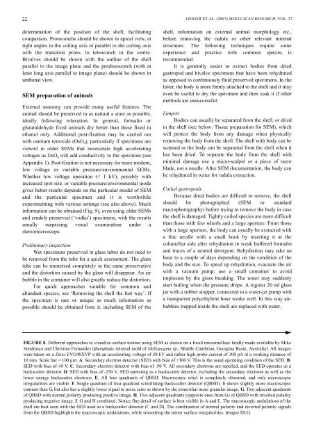
FIGURE 8. Different approaches to visualize surface texture using SEM as shown on a fossil micromollusc kindly made available by Mike<br />
Vendrasco and Christine Fernandez (phosphatic internal mold of Mellopegma sp., Middle Cambrian, Georgina Basin, Australia). All images<br />
were taken on a Zeiss EVO40XVP with an accelerating voltage of 20 kV and rather high probe current of 300 pA at a working distance of<br />
10 mm. Scale bar = 100 µm. A. Secondary electron detector (SED) with bias of +300 V. This is the usual operating condition of the SED. B.<br />
SED with bias of ±0 V. C. Secondary electron detector with bias of -50 V. All secondary electrons are repelled, and the SED operates as a<br />
backscatter detector. D. SED with bias of -250 V. SED operating as a backscatter detector, excluding the secondary electrons as well as the<br />
lower energy backscatter electrons. E. All four quadrants of QBSD. Macroscopic relief is completely obscured, and only microscopic<br />
irregularities are visible. F. Single quadrant of four quadrant scintillating backscatter detector (QBSD). It shows slightly more macroscopic<br />
contrast than G, but also has a slightly lower signal to noise ratio as shown by the somewhat more granular image. G. Two adjacent quadrants<br />
of QBSD with normal polarity producing positive image. H. Two adjacent quadrants (opposite ones from G) of QBSD with inverted polarity<br />
producing negative image. I. G and H combined. Notice fine detail of surface is best visible in A and E. The macroscopic undulations of the<br />
shell are best seen with the SED used as a backscatter detector (C and D), The combination of normal polarity and inverted polarity signals<br />
from the QBSD highlights the macroscopic undulations, while smoothing the minor surface irregularities. Images DLG.