Molluscan Research: Techniques for collecting, handling, preparing ...
Molluscan Research: Techniques for collecting, handling, preparing ...
Molluscan Research: Techniques for collecting, handling, preparing ...

You also want an ePaper? Increase the reach of your titles
YUMPU automatically turns print PDFs into web optimized ePapers that Google loves.
30<br />
thoroughly, preferably overnight in an incubator. When<br />
transferring radulae onto the cover slip, be careful to<br />
prevent water getting under the cover slip as it will<br />
unstick it.<br />
The glass cover slip is released from the histo-slide base<br />
by applying some water to the edge of the cover slip.<br />
Capillary <strong>for</strong>ces will pull the water under the cover slip<br />
and dissolve the glue. The loose cover slip is then<br />
mounted on a SEM stub with new glue.<br />
• Rhipidoglossate (Vetigastropoda) and docoglossate<br />
(Patellogastropoda, Polyplacophora) radulae have<br />
overlapping lateral and marginal teeth, which obscure<br />
other teeth. To mount these types of radulae, glue some<br />
thin pieces of wire, or hair, or needles of a diameter<br />
close to the width of the radula and cover them with<br />
glue (Fig. 12A, B). Cover slips may also be prepared<br />
with a series of wires etc. of different widths. Radulae<br />
can then be mounted longitudinally, on top of these<br />
wires with the marginal teeth bent outwards and<br />
downwards. This af<strong>for</strong>ds a better view than mounting<br />
radulae across the wire (e.g., Strasoldo 1991), but<br />
requires some practice. Breaking up a flat-mounted<br />
radula will also give the necessary data.<br />
The orientation of a mounted radula can be doublechecked<br />
under a light microscope with a 25 or 40x objective<br />
although care is needed as the working distance is only 1–0.2<br />
mm, depending on the lens. For radulae mounted on glass<br />
slides, commonly available transmitted light microscopes<br />
can be employed. Stub mounted radulae can be checked with<br />
compound microscopes equipped <strong>for</strong> (or improvised) epiillumination<br />
or with high power stereomicroscopes.<br />
A radula that has been accidentally improperly mounted<br />
can often be released from the mounting surface.<br />
• For PVA glue, add water with a paint brush or a fine<br />
pipette, soak the radula, remove and remount it. Do not<br />
disturb the glue excessively, as it may invade the radula.<br />
• If mounted on double-sided carbon tabs or tape, a<br />
radula can be released by soaking it in a large drop of<br />
water <strong>for</strong> a minute and peeling it off the carbon tab from<br />
one end. Then, the radula may be remounted and dried<br />
again. Once the radula has been sputter coated and<br />
viewed in the SEM, the radula is more firmly stuck to<br />
the carbon tab. Water will usually not be sufficient to<br />
release the radula, but ethanol usually works. Coated<br />
GEIGER ET AL. (2007) MOLLUSCAN RESEARCH, VOL. 27<br />
radulae are usually stiffer and more brittle than fresh<br />
ones.<br />
Very small specimens<br />
The following variation of the method above has been<br />
used <strong>for</strong> very small radulae (e.g., those of cimids with a<br />
length of ca 60 µm) or post-larval gastropods. Soak the<br />
specimen in a small quantity of distilled water: 1–6 drops<br />
with a fine pipette in the depression slide or 1/3 or less of the<br />
depth of the solid watch glass. Dissolve 1/4 of a tablet of<br />
KOH to 50 µl (= 1–2 drops) of water. The tablets can be<br />
readily split using a small stainless wire cutter. Heat the<br />
solution in the incubator at 50°C <strong>for</strong> 15 minutes or a little<br />
more. Usually the specimen will not dissolve but remain in a<br />
lump of clarified tissues. Transfer the lump with a needle<br />
(bent 90°), or a pair of <strong>for</strong>ceps (an inferior method because<br />
more fluid is transferred) to a drop of distilled water on a<br />
cover-slip. Usually the lump of tissue will dissolve in a<br />
fraction of a second. Add a drop of distilled water, close to,<br />
but separated from, the point where the specimen was<br />
dissolved and pull the radula over to this without allowing<br />
the two drops to merge. Remove the dirty water with a small<br />
piece of lint free tissue curled around the tips of a pair of<br />
<strong>for</strong>ceps and pinched in position by a little rubber band<br />
around the upper part of the <strong>for</strong>ceps. Wash the radula a<br />
couple of times more—the radula should now be in very<br />
clean water.<br />
Manipulation of radula<br />
Manipulation techniques vary with the mounting<br />
surface chosen. Mark the position of the radulae, because<br />
they are often easier to spot with a light microscope than in<br />
the SEM.<br />
For carbon tabs, the radula can be manipulated in a<br />
small (preferably distilled) water drop using a pair of fine<br />
tungsten needles. The water will evaporate at room<br />
temperature in one to two minutes (ethanol evaporates too<br />
fast <strong>for</strong> many small radulae, although it works very well with<br />
larger radulae and has less surface tension than water [but see<br />
remarks on some vetigastropod radulae above]). The surface<br />
tension of the water will help in flattening the radula along<br />
its long axis. At the point when the radula is still moist,<br />
but when there is no free water around the radula (a period of<br />
about two to three seconds), gently spread the radula out with<br />
the needles. The outer rows of radular teeth tend to fold<br />
over the central field when they dry, there<strong>for</strong>e it is important<br />
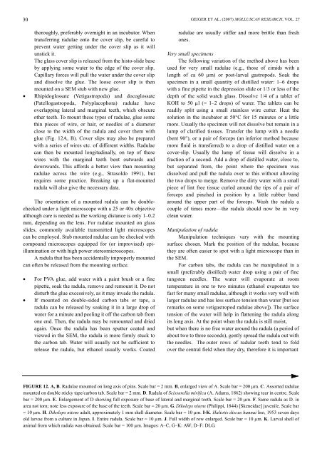
FIGURE 12. A, B. Radulae mounted on long axis of pins. Scale bar = 2 mm. B, enlarged view of A. Scale bar = 200 µm. C. Assorted radulae<br />
mounted on double sticky tape/carbon tab. Scale bar = 2 mm. D. Radula of Scissurella mirifica (A. Adams, 1862) showing tear in centre. Scale<br />
bar = 200 µm. E. Enlargement of D showing full exposure of base of lateral and marginal teeth. Scale bar = 20 µm. F. Same radula as D. in<br />
area not torn; note less exposure of the base of the teeth. Scale bar = 20 µm. G. Dikoleps nitens (Philippi, 1844) [Skeneidae] juvenile. Scale bar<br />
= 10 µm. H. Dikoleps nitens adult, approximately 1 mm shell diameter. Scale bar = 10 µm. I-K. Haliotis discus hannai Ino, 1953 seven days<br />
old larvae from a culture in Japan. I. Entire radula. Scale bar = 10 µm. J. Full width of row enlarged. Scale bar = 10 µm. K. Larval shell of<br />
animal from which radula was obtained. Scale bar = 100 µm. Images: A–C, G–K: AW; D–F: DLG.