Molluscan Research: Techniques for collecting, handling, preparing ...
Molluscan Research: Techniques for collecting, handling, preparing ...
Molluscan Research: Techniques for collecting, handling, preparing ...

Create successful ePaper yourself
Turn your PDF publications into a flip-book with our unique Google optimized e-Paper software.
26<br />
Extraction of radulae from micromolluscs<br />
The radulae of micromolluscs can be very small, often<br />
making manipulation daunting. However, with some practice<br />
and patience, radulae a fraction of a millimetre long can<br />
routinely be successfully mounted. If possible, use an adult<br />
specimen unless an ontogenetic study is specifically carried<br />
out, as the radulae of many species change morphology with<br />
age (Warén 1990). In some cases so-called generic characters<br />
are obtainable only from adult radulae.<br />
It is recommended that specimens used <strong>for</strong> radular<br />
preparation should be photographed prior to radular<br />
extraction, especially if there is any doubt as to identity. The<br />
shell may be destroyed when attempting to remove the body<br />
and, if chemical treatment is used, tissue-dissolving agents<br />
contribute to the deterioration of the shell. In some cases,<br />
species-level identification requires the observation of<br />
minute details such as protoconch microsculpture that cannot<br />
be observed with a light microscope and necessitates the use<br />
of SEM.<br />
While the radula can be dissected from larger<br />
microgastropods, there is a danger of damaging it. A safer<br />
method is to dissolve the buccal mass or even the entire<br />
animal.<br />
There are a number of methods used <strong>for</strong> dissolving the<br />
tissue surrounding the radula. The simplest and quickest<br />
methods can be used <strong>for</strong> most gastropods. Gentler, more time<br />
consuming and more complicated methods may be necessary<br />
<strong>for</strong> some of the groups with delicate radulae or radular<br />
membranes. The latter include:<br />
• Patellogastropoda. Damaged by strongly alkaline<br />
agents; mineralised cusps fall off and the remaining<br />
parts are partly dissolved.<br />
• Monoplacophora. Teeth damaged by strong alkali.<br />
GEIGER ET AL. (2007) MOLLUSCAN RESEARCH, VOL. 27<br />
• Lepetelloidea. Teeth may get distorted and crack in<br />
strong KOH.<br />
• Vetigastropoda.<br />
o In some with thin and slender teeth (e.g.,<br />
calliostomatids, trochaclids), the teeth become<br />
softer and tend to stick together. These should,<br />
after rinsing and cleaning, be soaked in 50%<br />
ethanol and mounted in at least 80% ethanol, to<br />
reduce the risk of the teeth sticking together. In<br />
ethanol the teeth become stiffer and there is less<br />
surface tension.<br />
o In some fissurellids (Cosmetalepas) the teeth fall<br />
off the radular membrane when treated with strong<br />
alkali.<br />
We advise against trying to dissolve the animal inside<br />
the shell. Strong NaOH or KOH will damage the organic<br />
matrix in the shell (Strasoldo 1991) and the remaining<br />
hydroxide will react with aerial carbon dioxide, to <strong>for</strong>m a<br />
crystalline or powdery coating that cannot be removed (Fig.<br />
10). However, with some sturdy gastropods, such as<br />
marginellids, no adverse effects have been reported (Coovert<br />
and Coovert 1987) and short periods of maceration of tissue<br />
inside the shell can be tried. Shells of more fragile<br />
gastropods, such as scissurellids, will break when sonicated<br />
after such treatment, whereas they are stable be<strong>for</strong>e radular<br />
extraction. Shells exposed to tissue dissolving agents also<br />
deteriorate over time and can be completely broken down in<br />
as little as 10 years, while enzymes in detergents can destroy<br />
a shell in a few hours, due to the low pH. Proteinase K,<br />
commonly used in DNA extraction from tissues, is most<br />
destructive to the shell. Whereas hydroxide treatment usually<br />
leaves a recognisable shell behind, proteinase K will<br />
fragment shells.<br />
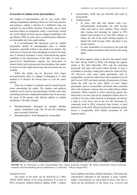
FIGURE 10. A. Protoconch of shell (Scissurellidae) after sodium hydroxide treatment. B. Similar protoconch without hydroxide<br />
treatment. Note the tunnelling and recrystallisation in A. Scale bars = 100 µm. Images DLG.<br />
Standard method<br />
The tissue of the body can be dissolved in 5–40%<br />
NaOH, KOH, bleach, or by using proteinase K as part of<br />
DNA extraction. Sodium dodecyl sulphate (SDS = sodium<br />
lauryl sulphate) provides a further alternative. The hydroxide<br />
concentration indicated in the literature is quite variable;<br />
higher concentrations are advocated by those who like to<br />
speed up the dissolution of the tissue (e.g., Coovert and