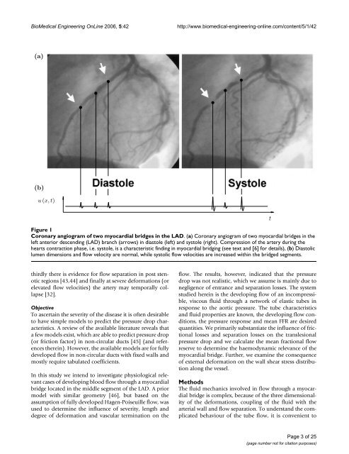
BioMedical Engineering OnLine 2006, 5:42http://www.biomedical-engineering-online.com/content/5/1/42BackgroundThe incomplete understanding of <strong>the</strong> pathophysiologyand clinical relevance of myocardial bridges has been <strong>the</strong>subject of debate for <strong>the</strong> last quarter century. An overviewof physiological relevant mechanisms of myocardialbridging, <strong>the</strong> current diagnostic <strong>to</strong>ols and treatment strategiesare found in [1-8]. Despite extensive studies on thissubject <strong>the</strong>re is no consensus on its clinical significance <strong>to</strong>myocardial ischaemia or angina pec<strong>to</strong>ris.A variety of models concerned with arterial stenoses [9-12] and series of stenoses [13-15] are found in <strong>the</strong> literature.Theoretical studies have been done <strong>to</strong> predict <strong>the</strong>location of maximum wall shear stress [16-18] and <strong>the</strong>extent of flow separation located distal <strong>to</strong> fixed stenoses[19-21]. There are a few models, which discuss flow in atime dependent two-dimensional flow geometry [22-25].These models assume rigid walls and are mainly focusedon vortex formation and <strong>the</strong> wall shear stress distribution.However, [26] discussed <strong>the</strong> extent of <strong>the</strong> separation zonein a one-dimensional empirical parameter model using<strong>the</strong> concept of dividing streamline. They found goodagreement with experiments in two-dimensional (partly)flexible indented channels [27].The interesting dynamic phenomena of collapsible tubesare discussed in [28-31]. When <strong>the</strong> tube wall is partiallycollapsed strong oscillations may occur, even understeady flow conditions. The non-linear coupling between<strong>the</strong> fluid pressure and tube wall deformation can produceconditions in which high-grade stenoses may collapse[32]. We note that in <strong>the</strong> late sys<strong>to</strong>le <strong>the</strong> compression of<strong>the</strong> artery in a myocardial bridge may cause conditionswhere <strong>the</strong> vessel is entirely closed and where <strong>the</strong> flow limitingeffect during re-opening becomes significant.Clinical situationUnder normal circumstances, coronary arteries havediameters large enough <strong>to</strong> transport sufficient amounts ofoxygen <strong>to</strong> myocardial cells. Increases in myocardial oxygendemand, e.g. during exercise, are met by increases incoronary artery blood flow because – unlike in manyo<strong>the</strong>r organs – extraction of oxygen from blood cannot beincreased. This is in part mediated by increases in diametersof small intra-myocardial arteries. The large proximal(epicardial) coronary arteries contribute only a small fractionof <strong>to</strong>tal vascular resistance and show little variation indiameter during <strong>the</strong> cardiac cycle in any given metabolicsteady state. Under maximum arteriolar vasodilation, <strong>the</strong>resistance imposed by <strong>the</strong> myocardial bed is minimal andblood flow is proportional <strong>to</strong> <strong>the</strong> driving pressure.The most common cause of an impaired ability <strong>to</strong> matchoxygen supply and demand is coronary a<strong>the</strong>rosclerosis, adisease that eventually leads <strong>to</strong> fixed coronary arterylumen narrowing, impaired coronary blood flow andpotentially myocardial infarction. However, some peoplepresent with chest pain caused by phasic lumen obstructiondue <strong>to</strong> myocardial bridging, first mentioned by Reymanin 1737 [33]. In this ana<strong>to</strong>mic variant, a coronaryartery segment courses underneath myocardial fibresresulting in vessel compression during sys<strong>to</strong>le, i.e. <strong>the</strong>myocardial contraction phase [6]. Myocardial bridges aremost commonly found in <strong>the</strong> mid LAD, 1 mm <strong>to</strong> 10 mmbelow <strong>the</strong> surface of <strong>the</strong> myocardium with typical lengthof 10 mm <strong>to</strong> 30 mm. An angiogram of two myocardialbridges in series shown in Figure 1(a).Although coronary blood flow occurs predominantly duringdias<strong>to</strong>le, i.e. <strong>the</strong> filling phase of <strong>the</strong> hearts chambers,<strong>to</strong>tal blood flow may none<strong>the</strong>less be reduced partlybecause vascular relaxation may extend significantly in<strong>to</strong>dias<strong>to</strong>le, <strong>the</strong> myocardial relaxation phase. Within <strong>the</strong>bridged segments permanent diameter reductions of 22 –58% were found during dias<strong>to</strong>le, while in sys<strong>to</strong>le <strong>the</strong>diameters were reduced by 70 – 95% [5]. A schematicdrawing of <strong>the</strong> increased flow velocities (cm/s) during sys<strong>to</strong>le(31.5 within versus 17.3 proximal and 15.2 distal) isgiven in Figure 1(b).From a medical point of view coronary angiography islimited in its ability <strong>to</strong> determine <strong>the</strong> physiologic significanceof coronary stenosis [34,35]. As a result, intracoronaryphysiologic measurement of myocardial fractionalflow reserve was introduced and has proven <strong>to</strong> be a reliable<strong>method</strong> for determining <strong>the</strong> functional severity of coronarystenosis. Previous studies have shown that <strong>the</strong> cu<strong>to</strong>ffvalue of 0.75 reliably detects ischaemia-producinglesions for patients with moderate epicardial coronary stenosis[36]. The assessment of <strong>the</strong> FFR is independent ofchanges in systemic blood pressure, heart rate, or myocardialcontractility and is highly reproducible [37]. The concep<strong>to</strong>f coronary pressure-derived FFR has beenextensively studied [13,38-40], clinically validated [41]and was found <strong>to</strong> be very useful in identifying patientswith multi-vessel disease [42], who might benefit fromca<strong>the</strong>ter-based treatment instead of surgical revascularisation.As in [38], we have defined <strong>the</strong> pressure derived FFRas <strong>the</strong> ratio between <strong>the</strong> pressures measured distal <strong>to</strong> andproximal <strong>to</strong> <strong>the</strong> myocardial bridge during maximal hyperaemia.The exact locations of pressure measurement aregiven later in <strong>the</strong> text.In summary myocardial bridges are characterised by aphasic sys<strong>to</strong>lic vessel compression with a persistentdias<strong>to</strong>lic diameter reduction, increased blood flow velocities,retrograde flow, and a reduced flow reserve [5]. Theunderlying mechanisms are fourfold. Firstly <strong>the</strong> discontinuitycauses wave reflections, secondly <strong>the</strong> dynamicreduction of <strong>the</strong> vessel diameter produces secondary flow,Page 2 of 25(page number not for citation purposes)
BioMedical Engineering OnLine 2006, 5:42http://www.biomedical-engineering-online.com/content/5/1/42(a)(b)u (x, t)Coronary Figure 1 angiogram of two myocardial bridges in <strong>the</strong> LADCoronary angiogram of two myocardial bridges in <strong>the</strong> LAD. (a) Coronary angiogram of two myocardial bridges in <strong>the</strong>left anterior descending (LAD) branch (arrows) in dias<strong>to</strong>le (left) and sys<strong>to</strong>le (right). Compression of <strong>the</strong> artery during <strong>the</strong>hearts contraction phase, i.e. sys<strong>to</strong>le, is a characteristic finding in myocardial bridging (see text and [6] for details), (b) Dias<strong>to</strong>liclumen dimensions and flow velocity are normal, while sys<strong>to</strong>lic flow velocities are increased within <strong>the</strong> bridged segments.tthirdly <strong>the</strong>re is evidence for flow separation in post stenoticregions [43,44] and finally at severe deformations (orelevated flow velocities) <strong>the</strong> artery may temporally collapse[32].ObjectiveTo ascertain <strong>the</strong> severity of <strong>the</strong> disease it is often desirable<strong>to</strong> have simple models <strong>to</strong> predict <strong>the</strong> pressure drop characteristics.A review of <strong>the</strong> available literature reveals thata few models exist, which are able <strong>to</strong> predict pressure drop(or friction fac<strong>to</strong>r) in non-circular ducts [45] (and references<strong>the</strong>rein). However, <strong>the</strong> available models are for fullydeveloped flow in non-circular ducts with fixed walls andmostly require tabulated coefficients.In this study we intend <strong>to</strong> investigate physiological relevantcases of developing blood flow through a myocardialbridge located in <strong>the</strong> middle segment of <strong>the</strong> LAD. A priormodel with similar geometry [46], but based on <strong>the</strong>assumption of fully developed Hagen-Poiseuille flow, wasused <strong>to</strong> determine <strong>the</strong> influence of severity, length anddegree of deformation and vascular termination on <strong>the</strong>flow. The results, however, indicated that <strong>the</strong> pressuredrop was not realistic, which we assume is mainly due <strong>to</strong>negligence of entrance and separation losses. The systemstudied herein is <strong>the</strong> developing flow of an incompressible,viscous fluid through a network of elastic tubes inresponse <strong>to</strong> <strong>the</strong> aortic pressure. The tube characteristicsand fluid properties are known, <strong>the</strong> developing flow conditions,<strong>the</strong> pressure response and mean FFR are desiredquantities. We primarily substantiate <strong>the</strong> influence of frictionallosses and separation losses on <strong>the</strong> translesionalpressure drop and we <strong>calculate</strong> <strong>the</strong> mean fractional flowreserve <strong>to</strong> determine <strong>the</strong> haemodynamic relevance of <strong>the</strong>myocardial bridge. Fur<strong>the</strong>r, we examine <strong>the</strong> consequenceof external deformation on <strong>the</strong> wall shear stress distributionalong <strong>the</strong> vessel.MethodsThe fluid mechanics involved in flow through a myocardialbridge is complex, because of <strong>the</strong> three dimensionalityof <strong>the</strong> deformations, coupling of <strong>the</strong> fluid with <strong>the</strong>arterial wall and flow separation. To understand <strong>the</strong> complicatedbehaviour of <strong>the</strong> tube flow, it is convenient <strong>to</strong>Page 3 of 25(page number not for citation purposes)
- Page 1: BioMedical Engineering OnLineBioMed
- Page 6 and 7: BioMedical Engineering OnLine 2006,
- Page 8 and 9: BioMedical Engineering OnLine 2006,
- Page 10 and 11: BioMedical Engineering OnLine 2006,
- Page 12: BioMedical Engineering OnLine 2006,
- Page 15 and 16: BioMedical Engineering OnLine 2006,
- Page 17 and 18: BioMedical Engineering OnLine 2006,
- Page 19 and 20: BioMedical Engineering OnLine 2006,
- Page 21 and 22: BioMedical Engineering OnLine 2006,
- Page 23 and 24: BioMedical Engineering OnLine 2006,
- Page 25: BioMedical Engineering OnLine 2006,