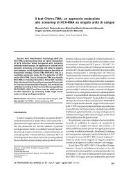
Flegel W AFigure 4 - Category DVI as a result of gene conversion. The two RH genes lie on their chromosome pointing in opposite directions(i.e., a cluster). When the chromosome folds, the two RH genes are adjacent, now pointing in the same direction. Thisconfiguration allows gene conversion in cis, whereby a DNA segment is transferred from one gene to another. The middlesection of the RHD gene (yellow) is substituted by the corresponding homologous section of the RHCE gene (green) (A).This type of gene conversion is responsible for the RHD-CE(3-6)-D allele, which codes for the D category VI of themolecular type 3 (DVI type 3) (B). Exons 1 to 10 are drawn on both RH genes (C). Due to the contrary directions, theterminal exons of the two RH genes (RHD <strong>and</strong> RHCE exon 10) lie closest to each other. On the RHD gene, exons 3 to 6 aresubstituted by the homologous exons of the RHCE gene.Weak DIf an amino acid substitution is located within theerythrocyte membrane or the cytoplasm, this will result in aweak D phenotype (figure 2) 11 . Integration of the RhDprotein into the membrane will be hindered, leading toquantitative weakening of the D antigen. There is usuallyno qualitative change, <strong>and</strong> hence no anti-D immunization.The weak D type 1 is the commonest in Europe (Table I).DELA particularly weakly expressed D antigen is termedDEL (earlier Del), because it could only be demonstratedusing elution. In elution, antibodies are separated fromerythrocytes to demonstrate them in the eluate. Themolecular changes are more severe than those seen withweak D, considerably hindering but not completelypreventing integration into the cell membrane. All DELalleles are rare in Europe, but up to 30% of all apparently Dnegative individuals in East Asia are bearers of the DELallele RHD (K409K) 10,12 .The C/c <strong>and</strong> E/e antigensThe clinically important Rhesus antigens C, c, E <strong>and</strong> eare the result of RhCE protein changes at only five aminoacid locations (figure 2). Antigens are termed antithetical ifa protein can present only one of them. They are causedby protein polymorphisms. Often there are two variants ofa protein, which differ at only one amino acid location,such as the Rhesus antigens E <strong>and</strong> e. RHCE alleles showingthe amino acid proline at position 226 express the E antigen,whereas RHCE alleles showing the amino acid alanine atthis position express the e antigen (Table I) 1 . Similardifferences between two RHCE alleles account for theantithetical C <strong>and</strong> c antigens. The antigen pairs C/c <strong>and</strong>E/e are not antithetical, however, because they result fromsubstitutions at different locations. The four possiblecombinations occur at different frequencies (amongEuropeans: Ce > ce > cE > CE) <strong>and</strong> are inherited ashaplotypes.Clinical applicationsGenetic investigation, like all investigations in <strong>medicine</strong>,should only be carried out in the context of a clear aim 13 . Asfar as <strong>transfusion</strong>s are concerned, molecular biologicaltechniques already are being used to provide cost-effectiveanswers to a number of clinically important questions.Methods used include polymerase chain reaction (PCR)for gene amplification <strong>and</strong> subsequent identification byelectrophoresis, nucleotide sequencing <strong>and</strong> hybridizationon biochips 14 .Anti-D in patientsThe clinical problems encountered are caused by a smallnumber of RHD alleles. Patients usually show partial D, insome rare cases weak D immunized by normal D antigen.Since category VI (DVI) is the most important of these, theAuthors recommend using monoclonal anti-D antibodiesfor typing, which do not react with DVI 15,16 .54<strong>Blood</strong> Transfus 2007; 5: 50-57050-57_flegel.p65 5405/07/2007, 10.34
Genetics of Rhesus systemThis procedure was included in the Germanhaemotherapy guidelines in 1996 <strong>and</strong> has not been changedsince. DVI carriers are therefore deliberately typed as falsenegatives to prevent <strong>transfusion</strong>s with D positive blood<strong>and</strong> likely anti-D immunization 17 .After these precautions were built into the Germanguidelines, they also were adopted by other Europeancountries. Unlike partial D, no anti-D alloimmunization hasyet been reported for weak D type 1, 2 or 3 18 . From a clinicalperspective, it is helpful that this involves the commonestD alleles, which make up almost 90% of all weak D types inGermany 19 , since these patients can receive D positiveblood <strong>transfusion</strong>s <strong>and</strong> do not require D negative products.This procedure saves up to 5% of all D negativeerythrocyte products, since they can perfectly well bereplaced by D positive products 11 , thus avoidingbottlenecks in the supply of D negative blood products 20 .Pregnant women <strong>and</strong> anti-D prophylaxisPregnant women with weak D types 1 to 3 can also begiven D positive blood <strong>transfusion</strong>s, <strong>and</strong> require no anti-Dprophylaxis. Each year, a one-off genetic test helps avoidrepeated administration of anti-D to 3,500 pregnant womenin Germany alone (up to 5% of all D negative pregnancies),<strong>and</strong> with it, all possible side effects of this prophylaxis,which these women do not require. Up to 5% of all anti-Dinjections are therefore unnecessary.A one-off genetic test is more cost-effective thanrepeated administration of anti-D products. In order toimplement this approach, the guidelines for medical careduring pregnancy <strong>and</strong> after birth (motherhood guidelines),issued by the Federal Committee of Physicians <strong>and</strong> HealthInsurance Funds [Bundesausschuss der Ärzte undKrankenkassen], would need to be adapted accordingly 21 .All pregnant women with rare weak D types would receivethe necessary prophylaxis, which they would notautomatically receive under the current state ofhaemotherapy 5 <strong>and</strong> motherhood guidelines 21 .A foetus can be shown to be D positive bydemonstrating foetal DNA in the plasma of peripheralmaternal blood 22 . Anti-D prophylaxis is unnecessary if thefoetus is D-negative. This could save about 40% of all theanti-D prophylaxis currently given during pregnancy. Thismethod was developed in countries bordering on Germany,where intensive efforts are under way to implement thisapproach to genetic diagnosis 23 .Prenatal diagnosisIf the foetus needs to be tested for D antigen,amniocentesis or sampling from the trophoblast is themethod of choice 14 . Cordocentesis is no longer performed.As already mentioned, maternal plasma may be able to beused in future.Having a child <strong>and</strong> anti-D antibodiesIf the father is heterozygous for the RHD deletion, thereis a 50% chance of the foetus being D-negative, in whichcase the pregnancy essentially free of any haematologicalrisk. If the father is homozygous for the RHD gene, thefoetus will definitely inherit the D antigen, which couldinfluence the couple's decision on whether to have a childor not.For several decades, it was impossible to determinewhether an individual is heterozygous or homozygous forRHD because serological methods are unsuitable. Withthe advent of the genetic diagnosis of the hybrid Rhesusbox, however, the possibilities have been exp<strong>and</strong>edconsiderably 9 . If the father is D-positive, it is now sufficientto test him for the RHD deletion.Use in other diseasesIf the st<strong>and</strong>ard serological methods fail, geneticdiagnosis is the method of choice for a reliable blood grouptyping of patients after a <strong>transfusion</strong> <strong>and</strong> those with autooralloimmune haematological anaemias. Althoughtransfused leucocytes can under certain circumstancespersist for years, they will not interfere with routine geneticdiagnosis.<strong>Blood</strong> donorsAppropriate investigation for the RHD gene canidentify apparently D negative donors, who in reality areweak D or DEL, thus ensuring that their blood will be givenonly to D positive recipients 18 . Without genetic diagnostics,D negative blood <strong>transfusion</strong> recipients will continue to beimmunized by the D antigen contained in such blood 24-27 .Donors who so far were misidentified as being Dnegative <strong>and</strong> whose erythrocytes are D-/D+ chimeras cannow be identified correctly. Lifelong chimerism can resultfrom monochorionic twin pregnancies. Any <strong>transfusion</strong>from donor sources such as these can result in anti-Dimmunization, because they also contain several millilitresof erythrocytes with a perfectly normal D positivephenotype. This D positive blood can only be detectedusing genetic investigation, not with routine serologicalmethods 10,27 . Any case of anti-D immunization is ofconsiderable clinical importance for girls <strong>and</strong> women ofreproductive age. In the case of a D positive pregnancy,<strong>Blood</strong> Transfus 2007; 5:50-5755050-57_flegel.p65 5505/07/2007, 10.34