Contents - Medicinski Fakultet u Sarajevu - University of Sarajevo
Contents - Medicinski Fakultet u Sarajevu - University of Sarajevo
Contents - Medicinski Fakultet u Sarajevu - University of Sarajevo
- No tags were found...

Create successful ePaper yourself
Turn your PDF publications into a flip-book with our unique Google optimized e-Paper software.
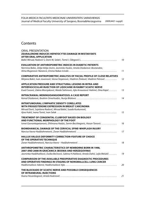
FOLIA MEDICA FACULTATIS MEDICINAE UNIVERSITATIS SARAEVIENSISJournal <strong>of</strong> Medical Faculty <strong>University</strong> <strong>of</strong> <strong>Sarajevo</strong>, Bosnia&Herzegovina2009;44(1-suppl)<strong>Contents</strong>Oral PresentationZEARALENONE INDUCES HEPATOCYTES DAMAGE IN WISTAR RATSAFTER ORAL APPLICATIONBabić Mirsad, Radović S, Dorić M, Selak I, Tomić I, Ožegović L. . . . . . . . . . . . . . . . . . . . . . . . . . . . . . . . . . . . . 10EVALUATION OF ANTHROPOMETRIC INDICES IN DIABETIC PATIENTSNermina Babic, Zelija Velija-Asimi, Jasminko Huskic, Amela Dizdarevic-Bostandzic,Alma Mujanovic-Narancic, Emina Nakas-Icindic . . . . . . . . . . . . . . . . . . . . . . . . . . . . . . . . . . . . . . . . . . . . . . . . . 11COMPARATIVE ANTROPOMETRIC ANALYSIS OF FACIAL PROFILE OF CLOSE RELATIVESMirjana Bakić, Ivan Jovanović, Vesna Stojanovic, Vladimir Živković, Vladimir Petrović . . . . . . . . . . . . 12APPLICATION PRESSURE AND STRUCTURAL LESIONS IN INTRA-ANDINTERFASCICULAR INJECTION OF LIDOCAINE IN RABBIT SCIATIC NERVEEsad Ćosović, Zakira Mornjaković, Maida Šahinovic, Ajla Arnautović-Halimić, Dina Kapić . . . . . . . . 13INTRACRANIAL MENINGIOANGIOMATOSIS: A CASE REPORTKemal Dizdarevic, Ibrahim Omerhodzic, Nurija Bilalovic . . . . . . . . . . . . . . . . . . . . . . . . . . . . . . . . . . . . . . . . . 14INTRATUMOURAL LYMPHATIC DENSITY CORELLATESWITH PROGESTERONE EXPRESSION IN BREAST CARCINOMAMirsad Dorić, Svjetlana Radović, Mirsad Babić, Suada Kuskunović,Ajna Hukić, Ivana Tomić, Ivan Selak . . . . . . . . . . . . . . . . . . . . . . . . . . . . . . . . . . . . . . . . . . . . . . . . . . . . . . . . . . . . . 15TREATMENT OF CONGENITAL CLUBFOOT BASED ON BIOLOGYAND FUNCTIONAL MORPHOLOGY OF THE FOOTIsmet Gavrankapetanovic, Ehlimana Hadzic, Semin Becirbegovic, Hasan Tanovic . . . . . . . . . . . . . . . . 16BIOMEHANICAL DAMAGE OF THE CERVICAL SPINE-WHIPLASH INJURYNarcisa Vavra-Hadžiahmetović, Zoran Hadžiahmetović . . . . . . . . . . . . . . . . . . . . . . . . . . . . . . . . . . . . . . . . . 17HALLUX VALGUS DEFORMITY CORRECTION-FEATURE OF CHOICEOF THE OPERATIVE TECHNIQUEZoran Hadžiahmetović, Narcisa Vavra – Hadžiahmetović . . . . . . . . . . . . . . . . . . . . . . . . . . . . . . . . . . . . . . . . 18ANTHROPOMETRIC CHARACTERISTICS OF NEWBORNS BORN IN 1998,2007 AND 2008 IN GRAČANICA (BOSNIA AND HERZEGOVINA)Jasminka Hadžihalilović, Duška Bećirović, Sabina H.Halilović, Amela Dahić, Lejla Mešalić . . . . . . . . 19Comparison <strong>of</strong> the available preoperative diagnostic proceduresand operative findings in staging <strong>of</strong> nonsmallcell lung cancerHadžismailovic Ademir, Hadžismailović Ajla . . . . . . . . . . . . . . . . . . . . . . . . . . . . . . . . . . . . . . . . . . . . . . . . . . . . . 20THE BLOCKADE OF SCIATIC NERVE AND POSSIBLE CONSEQUENCESOF INTRANEURAL INJECTIONSIlvana Hasanbegović, Amela Kulenović . . . . . . . . . . . . . . . . . . . . . . . . . . . . . . . . . . . . . . . . . . . . . . . . . . . . . . . . . . 21
MORPHOLOGICAL CHARACTERISTICS OF THE MYOCARDIIUMWITH COMPROMITATION OF CORONARY CIRCULATIONAida Hasanović, Zakira Mornjaković, Branko Pikula. . . . . . . . . . . . . . . . . . . . . . . . . . . . . . . . . . . . . . . . . . . . . . 22RAT KIDNEY DAMAGE AND BLOOD MYOGLOBIN LEVELAFTER ISOPROTERENOL ADMINISTRATIONHasić Sabaheta, Kiseljaković Emina, Ćosović Esad, Jadrić Radivoj,Winterhalter-Jadrić Mira, Mornjaković Zakira. . . . . . . . . . . . . . . . . . . . . . . . . . . . . . . . . . . . . . . . . . . . . . . . . . . . 23Hepatic steatosis as a predictor <strong>of</strong> early virological responseto pegilated interferon and ribavirin therapy in patientswith chronic hepatitis cAzra Husić-Selimović, Zora Vukobrat-Bijedić, Bisera Gogov. Srđan Gornjaković . . . . . . . . . . . . . . . . . . . 24BREAST CANCER–TODAY AND TOMORROWZerina Jašarevic. . . . . . . . . . . . . . . . . . . . . . . . . . . . . . . . . . . . . . . . . . . . . . . . . . . . . . . . . . . . . . . . . . . . . . . . . . . . . . . . . 25DIFFICULTIES IN TREATMENT OF RECTUM HYPOGANGLIONOSISWITH SYMPTOMS OF CHRONIC OBSTRUCTIONKenan Karavdić, Zlatan Zvizdić, Salahudin Dizdarević . . . . . . . . . . . . . . . . . . . . . . . . . . . . . . . . . . . . . . . . . . . 26EVALUTATION OF OXIGEN CONSUPTION OF HYBRIDE CLONES WITH POINTMUTATION ON tRNAser(UCN) OF MITOCHONDRIAL DNAEmina Kiseljaković, Sabaheta Hasić, Radivoj Jadrić, Zakira Mornjaković, Mira Winterhalter-Jadrić 27Histo-morphological characteristics <strong>of</strong> transplantationreactions to needle biopsies <strong>of</strong> kidneys at laboratory diagnosticspoliclinic– institute <strong>of</strong> pathology, university clinical centar tuzla,in the period 2000 – 2008Maja Konrad, Zinaida Karasalihović, Goran Šarkanović . . . . . . . . . . . . . . . . . . . . . . . . . . . . . . . . . . . . . . . . . . 28EXPRESSION OF CERTAIN MEMBERS OF BCL-2 FAMILY OF ONCOPROTEINSIN STREPTOZOTOCIN MODEL OF ALZHEIMER’S DISEASEKundurovic Z, Hasanagic S, Bilalovic N, Gavrankapetanovic F. . . . . . . . . . . . . . . . . . . . . . . . . . . . . . . . . . . . 29IMMUNOHISTOCHEMICAL EXPRESSION OF TIMP-1 IN BREAST CARCINOMA:CORRELATION WITH CLINICO-PATHOLOGICAL DATAS. Kuskunović, S. Radović, M. Dorić, A. Hukić, M.Babić, I. Tomić-Čuk, I. Selak . . . . . . . . . . . . . . . . . . . . . . . 30CYCLOOXYGENASE-2, p53 AND GLUCOSE TRANSPORTER-1 ARE MARKERSIN THE DEVELOPMENT OF GALLBLADDER CARCINOMASMateja Legan . . . . . . . . . . . . . . . . . . . . . . . . . . . . . . . . . . . . . . . . . . . . . . . . . . . . . . . . . . . . . . . . . . . . . . . . . . . . . . . . . . . 31VARIATIONS BRANCHING OF MAIN TRUNK OF LEFT CORONARY ARTERYAND IMPORTANCE OF HER DIAGONAL BRANCHAlmira Lujinović, Fehim Ovčina. . . . . . . . . . . . . . . . . . . . . . . . . . . . . . . . . . . . . . . . . . . . . . . . . . . . . . . . . . . . . . . . . . 32Histological status <strong>of</strong> the reproductive organs <strong>of</strong> female Wistarrats under the conditions <strong>of</strong> zearalenon intakeSejad Mačkić, Zakira Mornjaković, Ladislav Ožegović, Senad Prašović, Dina Kapić, Maida Šahinović 33TRANSFORAMINAL LIGAMENT AND FIRST LUMBAR INTERVERTEBRALFORAMEN-AN ANATOMIC REPORTD. L Marić, R. Gudović, D. M. Marić, B. Krstonošić, B. Srdić, S. Mijatov . . . . . . . . . . . . . . . . . . . . . . . . . . . . . . 34
CHROMOGRANINE A IMMUNOREACTIVITY IN THE MYENTERIC PLEXUS OF THEHUMAN FETAL DUODENUM IN THE THIRD AND THE FIFTH MONTH OF DEVELOPMENTNikolić I, Todorović V, Petrović V, Mitić D . . . . . . . . . . . . . . . . . . . . . . . . . . . . . . . . . . . . . . . . . . . . . . . . . . . . . . . . . 35MICROSATELLITE INSTABILITY IN COLORECTAL CANCERAmila Orucevic. . . . . . . . . . . . . . . . . . . . . . . . . . . . . . . . . . . . . . . . . . . . . . . . . . . . . . . . . . . . . . . . . . . . . . . . . . . . . . . . . . 36COMPARATIVE HISTOPATHOLOGY OF GASTRIC HELICOBACTER SPP. INFECTIONIN HUMANS, DOGS AND PIGSPrašović. S, Lalošević D, Alić. A, Beširović H, Kadrić M and Pašić Š . . . . . . . . . . . . . . . . . . . . . . . . . . . . . . . . . 37EXTRANODAL INTERDIGITATING DENDRITIC CELL SARCOMA: PATHOLOGIC ANDIMUNOHISTOCHEMICAL CHARACTERISTICS OF AN UNRECOGNIZED DISEASE ENTITYRadović S, Dorić M, Žujo H1, Babić M, Kuskunović S, Hukić A, Tomić I, Selak I. . . . . . . . . . . . . . . . . . . . . . 38STEREOLOGICAL ANALYSIS OF TERMINAL VILLI OF HUMAN PLACENTASASSOCIATED WITH EPH GESTOSISRamić S., Žigić Z., Marković S., Ramić N.. . . . . . . . . . . . . . . . . . . . . . . . . . . . . . . . . . . . . . . . . . . . . . . . . . . . . . . . . . 39DENTICLE- GENETICAL, STOMATOLOGICAL AND MEDICAL PROBLEM. CASE REPORTAmira Redžić, Aida Berisalić, Fehrudin Mucić. . . . . . . . . . . . . . . . . . . . . . . . . . . . . . . . . . . . . . . . . . . . . . . . . . . . . 40PARATHYREODECTOMY IN PATIENTS ON HEMODIALYSISResić H., Kapidžić A., Kukavica N., Šahović V., Čorić A., Avdić E., Mašnić F.. . . . . . . . . . . . . . . . . . . . . . . . . . 41DIFFERENTIAL DIAGNOSIS OF CLEAR CELL TUMORSAzra Sadikovic, Ermina Iljazovic, Goran Šarkanovic . . . . . . . . . . . . . . . . . . . . . . . . . . . . . . . . . . . . . . . . . . . . . . 42ASYMMETRY OF LIMBIC STRUCTURE AT PATINETS WITH COMPLEX PARTIAL ATTACSAida Sarač - Hadžihalilović, Amela Kulenović . . . . . . . . . . . . . . . . . . . . . . . . . . . . . . . . . . . . . . . . . . . . . . . . . . . . 43ESTIMATION OF HER2 GENE AMPLIFICATION BY CHROMOGENIC IN SITUHYBRIDIZATION (CISH) OF BREAST CARCINOMA WITH EQUIVOCAL EXPRESSIONOF HER2 PROTEIN ANALIZED BY IMMUNOHISTOCHEMICAL METHODGoran Šarkanović, Jasminka Mustedanagić-Mujanović, Azra Sadiković, Maja Konrad . . . . . . . . . . . . 44STEREOLOGY METHOD ON THE ULTRASTRUCTURAL LEVEL – EXAMPLEOF A BREAST DUCTAL EPITHELIUM ANALYSEAličelebić Selma. . . . . . . . . . . . . . . . . . . . . . . . . . . . . . . . . . . . . . . . . . . . . . . . . . . . . . . . . . . . . . . . . . . . . . . . . . . . . . . . . 45REVIEW OF THE SUPERNUMERARY KIDNEY ARTERIES BY THE METHODOF THE UNSELECTIVE ANGIOGRAPHY PER SELDINGERElvira Talović, Amela Kulenović, Alma Voljevica . . . . . . . . . . . . . . . . . . . . . . . . . . . . . . . . . . . . . . . . . . . . . . . . . . 46THE INFLUENCE OF SPORT ACTIVITIES ON THE LENGTH OF PREMENSTRUALPERIOD IN GIRLS FROM TUZLA CANTONRasima Tupkušić, Jasminka Hadžihalilović, Amela Begić, Rifet Terzić, Elmira Hajder. . . . . . . . . . . . . . . 47HEMODINAMIC CAPACITIES OF WILLIS RINGVoljevica A, Kulenović A . . . . . . . . . . . . . . . . . . . . . . . . . . . . . . . . . . . . . . . . . . . . . . . . . . . . . . . . . . . . . . . . . . . . . . . . . 48HISTOLOGICAL FINDINGS AND ALT LEVEL IN PATIENTS WITH CHRONIC HEPATITIS CZora Vukobrat-Bijedić, Azra Husić-Selimović, Bisera Gogov. . . . . . . . . . . . . . . . . . . . . . . . . . . . . . . . . . . . . . . 49
EFFECTS ON THE RAT AND RABBIT ORGANS FUNCTIONAND MORPHOLOGY AFTER MULTIPLE APPLICATION OF COMBINATIONOF ALPHA – ADRENOCORTICOTROPINE 1-13 AND METHIONINE-ENKEPHALINMijanovic M, Rakanovic-Todic M, Kusturica J, Kulo, A, Mulabegovic N, Becic F, Doric M, Babic M . . . . 64MORPHOLOGY OF HUMAN PERI-CORONARY ADIPOCYTESRancic G, Atanassova P, Chaldakov G, Petrovic V, Rancic I, Cvetanovic J, Vukadinovic M, Petrović A. 65MORPHOMETRIC CHARACTERISTICS OF NEPHRITIC SINUS DURINGTHE AGEING PROCESS IN HUMANVesna Stojanović, Ivan Jovanović, Slađana Ugrenović, Mirjana Bakić, Vladimir Živković,Vladimir Petrovic. . . . . . . . . . . . . . . . . . . . . . . . . . . . . . . . . . . . . . . . . . . . . . . . . . . . . . . . . . . . . . . . . . . . . . . . . . . . . . . . 66INFECTIONS OF HIP JOINT ENDOPROTHESESAdnana Talić-Tanović, Zoran Hadžiahmetović, Ismet Gavrankapetanović, Edib Jerlagić . . . . . . . . . . 67THE ACCUMULATION OF LIPOFUSCIN IN PIRAMIDAL CELLSOF ISOCORTEX AND ALOCORTEXŽivković Vladimir, Snežana Pavlović, Natalija Stefanović, Mirjana Bakić, Vesna Stojanović. . . . . . . . 68GENOTOXIC IMPACT OF WEAK MAGNETIC FIELDS ON MITOSISIN ALLIUM CEPA’S CELLSJasmin Musanovic, Zijad Muharemovic, Azra Metovic, Marijana Filipovska-Musanovic . . . . . . . . . . 69THE MORPHOLOGY AND PHYSICAL PROPERTIES OF CONDUCTINGPOLYMER POLYANILINE DOPED WITH SULFURICZijad Muharemovic, Izet Gazdic, Dinko Babić, Mirsad Babić. . . . . . . . . . . . . . . . . . . . . . . . . . . . . . . . . . . . . . 70
Editor-in-ChiefBakir MehićExecute EditorMensura KudumovićEditorial BoardNedžad MulabegovićDamir AganovićJasmin MušanovićMuzafer MujićSemra ČavaljugaNermin SarajlićAlmira Hadžović DžuvoSlavenka VobornikRadivoj JadrićLectorised byDubravko VaničekTechnical Editor and PrintSaVart <strong>Sarajevo</strong>DTPNarcis PozderacAdress <strong>of</strong> the Editorial board:71000 <strong>Sarajevo</strong>, Čekaluša 90Bosnia & HerzegovinaPhone: 00387 33 663 472Fax: 00387 33 203 670Published byFaculty <strong>of</strong> Medicine,<strong>University</strong> <strong>of</strong> <strong>Sarajevo</strong>www.mf.unsa.ba/foliaISSN 0352-9630EBSCO Publishing (EP) USAhttp://www.epnet.comPrinted on acid-free paper
Treći BH simpozijum“Morfologija u nauci i praksi“sa međunarodnim učešćem23. - 25. septembar 2009.<strong>Sarajevo</strong>, Bosna i HercegovinaPokroviteljiAkademija nauka i umjetnosti BIHKlinički centar Univerziteta u <strong>Sarajevu</strong>Ljekarska - Liječnička komora Kantona <strong>Sarajevo</strong>Generalni sponzoriBOSNALIJEKGlavni sponzorOLYMPUSSponzoriKlinički centar Univerziteta u <strong>Sarajevu</strong>Ljekarska – Liječnička komoraBERLIN - CHEMIEDIAMEDICDonatorOrdinacija dr Begeta
Oral presentationTreći BH simpozijum “Morfologija u nauci i praksi“sa međunarodnim učešćemOrganizator<strong>Medicinski</strong> fakultet Univerziteta u <strong>Sarajevu</strong>Institut za morfološke nauke i genetikuKatedra za patologijuPokroviteljiAkademija nauka i umjetnosti BIHKlinički centar Univerziteta u <strong>Sarajevu</strong>Ljekarska - Liječnička komora Kantona <strong>Sarajevo</strong>Organizacioni odborPočasni predsjednik: Ivan SelakPredsjednik: Svjetlana RadovićDopredsjenici: Zakira Mornjaković, Amela KulenovićČlanovi: Mirsad Dorić, Mirsad Babić, Ivana TomićSekretari: Suada Kuskunović, Ajna HukićLektor za engleski jezik: Ajna HukićBlagajnik: Pehlivanović RemzijaNaučni odborAličelebić Selma (<strong>Sarajevo</strong>, BIH), Arnautović Ibrahim (<strong>Sarajevo</strong>, BIH), Cvetko Erika (Ljubljana, Slovenija),Dinarević-Mesihović Senka (<strong>Sarajevo</strong>, BIH), Dizdarević Kemal (<strong>Sarajevo</strong>, BIH), Dorić Mirsad(<strong>Sarajevo</strong>, BIH), Gavrankapetanović Ismet (<strong>Sarajevo</strong>, BIH), Hadžiahmetović Zoran (<strong>Sarajevo</strong>, BIH),Ibrulj Slavica (<strong>Sarajevo</strong>, BIH), Jadrić Radivoj (<strong>Sarajevo</strong>, BIH), Jašarević Zerina (Austrija), KapidžićAdnan (<strong>Sarajevo</strong>, BIH), Kapur Eldan (<strong>Sarajevo</strong>, BIH), Kulenović Amela (<strong>Sarajevo</strong>, BIH), Legan Mateja(Ljubljana, Slovenija), Marić Dušica (Novi Sad, Srbija), Mijatov Ljiljana (Novi Sad, Srbija),Mornjaković Zakira (<strong>Sarajevo</strong>, BIH), Emina Nakaš-Ićindžić (<strong>Sarajevo</strong>, BIH), Nikolić Ivan (Niš, Srbija)Oručević Amila (USA), Petrović Aleksandar (Niš, Srbija), Radović Svjetlana (<strong>Sarajevo</strong>, BIH),Ravnik Dean (Ljubljana, Slovenija), Redžić Amira (<strong>Sarajevo</strong>, BIH), Sarajlić Nermin (<strong>Sarajevo</strong>, BIH),Suljić Enra (<strong>Sarajevo</strong>, BIH), Živković Vladimir (Niš, Srbija).Počasni odborArslanagić Edin (<strong>Sarajevo</strong>, BIH), Begeta Ibrahim (<strong>Sarajevo</strong>, BIH), Faletar Luka (Mostar, BIH),Gavrankapetanović Faris (<strong>Sarajevo</strong>, BIH), Lincender Lidija (<strong>Sarajevo</strong>, BIH), Mehić Bakir (<strong>Sarajevo</strong>, BIH),Mulabegović Nedžad (<strong>Sarajevo</strong>, BIH), Hajrija Raščić-Konjhodžić (<strong>Sarajevo</strong>, BIH).
Poštovane kolege,Izuzetno nam je zadovoljstvo da prisustvujete Trećem BH simpozijumu “Morfologija u nauci ipraksi“ sa međunarodnim učešćem, koji se održava u <strong>Sarajevu</strong> od 23. do 25. septembra 2009.godine. Simpozij, koji je utemeljen 2003. i održava se svake treće godine, ovaj put organizujeKatedra za patologiju Medicinskog fakulteta Univerziteta u <strong>Sarajevu</strong>.Cilj ovih susreta je promocija i poticaj naučnih i stručnih istraživanja, poticaj za novimsaznanjima u cilju postizanja viših standarda u medicinskoj praksi.Na Simpozijumu, uz eminentne predavače iz inozemstva, učestvuju i brojni stručnjaci iz oblastipredkliničkih i kliničkih medicinskih disciplina. Apstrakti svih pristiglih radova publikovani suu časopisu „FOLIA MEDICA“ koji izdaje <strong>Medicinski</strong> fakultet Univerziteta u <strong>Sarajevu</strong>.Ovogodišnji Simpozij organizuje se pod pokroviteljstvom Akademije nauka i umjetnostiBIH, Kliničkog centra Univerziteta u <strong>Sarajevu</strong> i Ljekarske - Liječničke komore Kantona <strong>Sarajevo</strong>,a bodovat će se po Pravilniku Ljekarske -Liječničke komore Kantona <strong>Sarajevo</strong>.Mjesto održavanja Simpozija je amfiteatar Medicinskog fakulteta u krugu Kliničkog centra Univerzitetau <strong>Sarajevu</strong> (Ulica Bolnička 25). Pored radnog, pripremljen je i bogat zabavni program.Želimo Vam ugodan boravak u <strong>Sarajevu</strong>, gradu tradicionalno dobrih i pažljivih domaćina.S poštovanjem,Svjetlana RadovićPredsjednik Organizacionog odboramensurak@yahoo.comDistinguished colleagues,It is an exceptional pleasure to invite you to participate Third BH symposium “Morphology inscience and practice“ with international participation that will take place in <strong>Sarajevo</strong>, 23 rd to 25 thSeptember, 2009. The Symposium was founded in 2003 and is being held every three years, thistime is organized by the Department <strong>of</strong> Pathology, Medical Faculty <strong>University</strong> <strong>of</strong> <strong>Sarajevo</strong>.The aim <strong>of</strong> this scientific meeting is promotion and stimulation <strong>of</strong> scientific and pr<strong>of</strong>essionalinvestigation, impulse to new knowledge in order to achieve higher standard in medicalpractice.Eminent lecturers from abroad as well as numerous pr<strong>of</strong>essional in area <strong>of</strong> preclinical andclinical medical will participate in work <strong>of</strong> the Symposium. All abstract are published in journal“FOLIA MEDICA” issued by the Medical Faculty <strong>University</strong> <strong>of</strong> <strong>Sarajevo</strong>.This year Symposium is organized under the auspices <strong>of</strong> Academy <strong>of</strong> Science and Art BIH,Clinical Center <strong>University</strong> <strong>of</strong> <strong>Sarajevo</strong> and Medical Chamber <strong>of</strong> <strong>Sarajevo</strong> Canton and is going to beawarded with points in accordance with the Rulebook <strong>of</strong> Medical Chamber <strong>of</strong> <strong>Sarajevo</strong> Canton.The Symposium is going to take place at the Amphitheatre <strong>of</strong> Medical Faculty located inthe Clinical Center <strong>University</strong> <strong>of</strong> <strong>Sarajevo</strong> (Bolnička street no.25). Besides work program weprepared various social events.We wish you a pleasant stay in <strong>Sarajevo</strong>, city known for traditionally good and consideratehosts.Sincerely yours,Svjetlana RadovićPresident <strong>of</strong> the Organizational Board
Oral presentationZEARALENONE INDUCES HEPATOCYTES DAMAGE IN WISTARRATS AFTER ORAL APPLICATIONBabić Mirsad 1 , Radović S 1 , Dorić M 1 , Selak I 1 , Tomić I 1 , Ožegović L 21Institute <strong>of</strong> pathology Medical Faculty <strong>of</strong> <strong>Sarajevo</strong>2Academy <strong>of</strong> Sciences and Arts <strong>of</strong> Bosnia andHerzegovinaE-mail: mirsadbabic@yahoo.comObjective: Mycotoxin zearalenone is a product <strong>of</strong> fungiFusarium species which has activity similar to estrogen.Aim was to investigate toxic effects <strong>of</strong> mycotoxin zearalenoneon hepatocytes <strong>of</strong> experimental Wistar rats followingoral application.Material and methods: We used 42 experimental Wistarrats and orally applied mycotoxin dissolved in oilin different concentrations, 0,5 mg , 2 mg and 4 mg perkilogram <strong>of</strong> body weight, in the three time periods -10, 20 and 30 days. The liver tissue fixed in bufferedformaldehyde. Paraffin slices, 4 microns thicknesswere stained with histochemical and immunochemicalmethods.Results: Examination proved focal necrosis with depletion<strong>of</strong> PAS positive material in hepatocytes cytoplasmat all groups which received different doses. The mostexpressive focal necrosis was in group who receiveddoses <strong>of</strong> 0,5 mg ( p=0,005).Conclusion: In rats liver zearalenone caused focalnecrosis <strong>of</strong> hepatocytes. The experiment didn’t provesigns non-parenchymatous liver damage at mycotoxinactivity in the three different doses and periods.Key words: zearalenone, hepatocytes, focal necrosis,Wistar rats.10
Oral presentationEVALUATION OF ANTHROPOMETRIC INDICESIN DIABETIC PATIENTSNermina Babic 1 , Zelija Velija-Asimi 2 , Jasminko Huskic 1 , Amela Dizdarevic-Bostandzic 2 , Alma Mujanovic-Narancic 2 , Emina Nakas-Icindic1Institute <strong>of</strong> Physiology and Biochemistry,Medical Faculty, <strong>University</strong> <strong>of</strong> <strong>Sarajevo</strong>, <strong>Sarajevo</strong>,Bosnia and Herzegovina2Clinics for Endocrinology, Diabetes Mellitus andMetabolic Diseases, <strong>University</strong> Clinical Center <strong>of</strong><strong>Sarajevo</strong>, <strong>Sarajevo</strong>, Bosnia and HerzegovinaE-mail: nerminab@yahoo.comIntroduction: Abdominal obesity is a significant riskfactor for cardiovascular diseases. The aim: To researcha relationship between an abdominal adiposity, assessedby various anthropometric indices (body massindex (BMI), waist circumference (WC), waist-to-hipratio (WHR), waist-to-height ratio (W/HtR)), withpulse pressure (PP) in diabetic patients.Methods: The study included 30 patients with diabetesmellitus Type 1 (DM1), 30 patients with diabetes mellitusType 2 (DM2) and 30 healthy subjects. Anthropometricmeasurements were made for all subjects,including body height, weight, waist and hip circumference.All subjects were measured blood pressure.Results: All examined anthropometric indices and PPwere statistically significantly higher in patients withDM2, compared with other groups (p
Oral presentationCOMPARATIVE ANTROPOMETRIC ANALYSISOF FACIAL PROFILE OF CLOSE RELATIVESMirjana Bakić, Ivan Jovanović, Vesna Stojanovic, Vladimir Živković,Vladimir PetrovićInstitute <strong>of</strong> anatomy, Faculty <strong>of</strong> Medicine, NišE-mail: bakic@medfak.ni.ac.rsIntroduction Proportional facial esthetics and optimalfunctional occlusion have always been considered majorgoals <strong>of</strong> orthodontic treatment. Aim Using angularphotogranometry we measured anthropometric parameters<strong>of</strong> s<strong>of</strong>t tissues <strong>of</strong> facial pr<strong>of</strong>ile.Material and methods Digital photos <strong>of</strong> 96 subjects,members <strong>of</strong> three generations <strong>of</strong> different families wereused during the study. Anthropometric parameterswere measured by means <strong>of</strong> Image J s<strong>of</strong>tware.Conclusion The results showed no significant differencesin facial convexity, maxillary arch, and nas<strong>of</strong>rontalangle, angle <strong>of</strong> the nasal dorsum and medial third <strong>of</strong> theface in members <strong>of</strong> different generations. The values <strong>of</strong>vertical height <strong>of</strong> the face and nasal angle <strong>of</strong> the youngestfamily members were similar to the values <strong>of</strong> oldermale family members, while the values <strong>of</strong> mandibularangle <strong>of</strong> the youngest family members were similar toolder female family members. The lower third <strong>of</strong> theface <strong>of</strong> the youngest female members was similar in sizeto older male family members, while in the youngestmale family members it was similar to that <strong>of</strong> older femalemembers. The values <strong>of</strong> nas<strong>of</strong>rontal angle showeddifferent trend. Therefore, angular photogranometrywould be a useful method <strong>of</strong> predicting the growth anddevelopment <strong>of</strong> some facial parts in children.Key words: anthropometry, facial pr<strong>of</strong>ile12
Oral presentationAPPLICATION PRESSURE AND STRUCTURAL LESIONSIN INTRA-AND INTERFASCICULAR INJECTION OF LIDOCAINEIN RABBIT SCIATIC NERVEEsad Ćosović, Zakira Mornjaković, Maida Šahinovic,Ajla Arnautović-Halimić, Dina KapićInstitute <strong>of</strong> Histology and Embryology,Medical Faculty, <strong>University</strong> <strong>of</strong> <strong>Sarajevo</strong>,Čekaluša 90, 71000 <strong>Sarajevo</strong>,Bosnia and Herzegovina,E-mail: ehcosovic@yahoo.comIntroduction: Injection injuries <strong>of</strong> peripheral nervesmay occur as a complication <strong>of</strong> locoregional anesthesia,whereby the extent <strong>of</strong> damage depends on injectionsite, needle type, characteristics and dosage <strong>of</strong> the localanesthetic, value <strong>of</strong> application pressure. Our aim wasto verify the difference between the intra- and interfascicularapplication <strong>of</strong> lidocaine.Materials and methods: Using an automated infusionpump, lidocaine was administrated bilaterally in sciaticnerves <strong>of</strong> 5 rabbits (4-ml dose, application speed 3ml/min). Pressure values were recorded by a manometerand analyzed using the BioBench 1.2 s<strong>of</strong>tware. Sevendays after the injection, at the site <strong>of</strong> administration,a nerve segment (1 cm) was excised in vivo and wasprepared for histological analysis.Results: The mean value <strong>of</strong> application pressure attainedduring intrafascicular application (55,97±22,85kPa) was significantly higher in comparison to thevalue <strong>of</strong> application pressure attained during interfascicularapplication (18,70±5,43 kPa), just like the extent<strong>of</strong> reactive changes <strong>of</strong> the entire nerve structure. Thesechanges included thickening and hypercellularity <strong>of</strong>the epineurium, disintegration <strong>of</strong> the perineurium anddamage primarily <strong>of</strong> the myelinated nerve fibers.Conclusion: Avoidance <strong>of</strong> high values <strong>of</strong> applicationpressure during locoregional anesthesia is a possiblefactor in the prevention <strong>of</strong> nerve damage.Key words: sciatic nerve, intraneural application, lidocaine,histological analysis, rabbit13
Oral presentationINTRACRANIAL MENINGIOANGIOMATOSIS: A CASE REPORTKemal Dizdarevic 1, Ibrahim Omerhodzic 1 , Nurija Bilalovic 21Department <strong>of</strong> Neurosurgery Clinical Centre<strong>University</strong> <strong>of</strong> <strong>Sarajevo</strong>, BiH2Institute for pathology, Clinical Centre<strong>University</strong> <strong>of</strong> <strong>Sarajevo</strong>, BiHE-mail: kemaldiz@bih.net.baObjective: Meningioangiomatosis is a rare, focal corticaldisorder <strong>of</strong> benign nature characterized by a proliferativeprocess. Potentially amenable to surgical cure,early recognition <strong>of</strong> this condition is a key to avoidingunnecessary alternative therapies. The first case <strong>of</strong> sporadicintracranial meningioangiomatosis diagnosedand microsurgically treated in Bosnia is presented. Presentation:A 13-year-old male, previously fit and havingno family history <strong>of</strong> note, presented with a 2-yearhistory <strong>of</strong> intractable seizures unresponsive to medication,associated with headache but no FND. Intervention:Keyhole craniotomy and an interhemisphericfrontal approach was performed and total resection <strong>of</strong>the lesion was carried out. The specimen was noted tobe <strong>of</strong> firm consistency with well-demarcated edges andminimal vascularity. The intraoperative impression <strong>of</strong>the lesion was that <strong>of</strong> a low-grade glioma. Light microscopyshowed highly cellular areas showing a storiformgrowth pattern and occasional cellular whorl pattern.There was complete effacement <strong>of</strong> cortical architecture.On H&E staining, spindle-shaped cells proliferating ina fascicular pattern were noted. Reticulin was abundantin these areas. In the background, multiple foci <strong>of</strong>psammoma bodies were noted. Conclusion: The patientremained seizure-free and <strong>of</strong>f antiepileptic medicationspostoperatively. There was no recurrence in the12-months follow up period.Key words: Meningioangiomatosis, aggressivemeningeoma.14
Oral presentationINTRATUMOURAL LYMPHATIC DENSITY CORELLATES WITHPROGESTERONE EXPRESSION IN BREAST CARCINOMAMirsad Dorić * , Svjetlana Radović, Mirsad Babić, Suada Kuskunović,Ajna Hukić, Ivana Tomić, Ivan SelakInstitute <strong>of</strong> Pathology, Faculty <strong>of</strong> Medicine,<strong>University</strong> <strong>of</strong> <strong>Sarajevo</strong>,Čekaluša 90, 71000 <strong>Sarajevo</strong>,Bosnia and HerzegovinaE-mail: mdoric@lsinter.netBackground: Controversy exists regarding the topography<strong>of</strong> lymph vessels in breast cancer, their usefulnessas prognostic factors and whether active lymphangiogenesisoccurs within the tumor. Goal was investigateto presence intratumoral lymphatic vessels, existence <strong>of</strong>lymphangiogenesis and their role in tumor dissemination.Materials and methods: We investigated lymphaticvessels in 75 specimens <strong>of</strong> invasive breast carcinoma byimmunostaining for D2 40. Distribution and density <strong>of</strong>lymphatics were assessed intratumoral and peritumoraland correlated with clinicopathological parameters.Endothelial proliferation in lymphatic vessels was analyzedby dual-color immunohistochemistry with D2 40and Ki 67 respectively.Results: We demonstrated existence <strong>of</strong> intratumorallymphatic vessels whose density compared to peritumoralones is significantly lower (p=0, 0001). Decrease<strong>of</strong> intra and peritumoral lymphatic vessels density(LVD) compared to fibrocystic breast disease was observed(p=0,002). Density <strong>of</strong> intratumoral lymphaticvessels correlated with amount <strong>of</strong> progesterone receptorsin primary tumor (p=0,036). Intratumoral lymphangiogenesiswas absent or minimal.Conclusion: High intratumoral LVD is a demonstrator<strong>of</strong> low destructive potential and vice versa, and role <strong>of</strong>lymphatics in tumor progression is passive.Key words: intratumoral lymphatic density, breast carcinoma,lymphangiogenesis, progesterone15
Oral presentationTREATMENT OF CONGENITAL CLUBFOOT BASED ON BIOLOGYAND FUNCTIONAL MORPHOLOGY OF THE FOOTIsmet Gavrankapetanovic, Ehlimana Hadzic, Semin Becirbegovic, Hasan TanovicClinic for Orthopedic and Traumatology,Clinical Center <strong>University</strong> <strong>of</strong> <strong>Sarajevo</strong>,Bosnia and HerzegovinaE-mail: ismetcap@ortotrauma.com.baIntroduction: To determine the role <strong>of</strong> non-operativetreatment by Ponseti management in children withcongenital clubfoot. Ponseti management is based onanatomical and histological studies. The studies unveiledthat collagen in the ligaments <strong>of</strong> clubfoot is easilystretched. Findings implicated that with manipulation<strong>of</strong> the foot and maintaining correction by cast andbraces, deformity could be fully reduced without any orwith minimal invasive surgery.Material and Methods: In our Clinic many kinds <strong>of</strong>posteromedial release <strong>of</strong> clubfoot were done. Since2006 all children were treated by Ponseti. During 2006-2008 we had observed 74 patients with congenital clubfoot.After a series <strong>of</strong> casting by Ponseti, all <strong>of</strong> themwere evaluated by Pirani Scoring to determine whetherthe tenotomy <strong>of</strong> Achill tendon is indicated. After thechildren begun to walk, the function <strong>of</strong> foot was evaluated.Results: Since 2006, 74 children with congenital clubfoot(10 day to 9 months) were treated by Ponseti. Afterevaluation by Pirani Scoring, in 64 cases tenotomy wasdone. In one case we had equines relapse and in onecase cavus deformity.Conclusion: This work illustrates the benefits <strong>of</strong> Ponsetimanagement <strong>of</strong> clubfoot regarding good function<strong>of</strong> the foot and lower percentage <strong>of</strong> relapses.Key words: clubfoot, Ponseti management, casting16
Oral presentationBIOMEHANICAL DAMAGE OF THE CERVICALSPINE-WHIPLASH INJURYNarcisa Vavra – Hadžiahmetović 1 , Zoran Hadžiahmetović 21Clinic for physical medicine and rehabilitation2Emergency Department<strong>Sarajevo</strong>, Bolnička 25, Bosnia and HerzegovinaE-mail: h.vemi@bih.net.baIntroduction: Whiplash, also called neck sprain orneck strain, is an injury to the s<strong>of</strong>t tissues <strong>of</strong> the neckcaused by biomechanical injury <strong>of</strong> the cervical spine.The exact injury mechanism that causes whiplash injuriesis unknown. The aim <strong>of</strong> this paper is to make rulesfor rehabilitation. Examines and Methods: 40 patientswith WNI were treated in Clinical center <strong>University</strong> <strong>of</strong><strong>Sarajevo</strong> (Clinic for physiotherapy and rehabilitationand in Clinic for urgent medicine) in period 2004/2008.They were divided in two groups – (G1 and G2)with20 patients in each group.G1 group was treated by s<strong>of</strong>tcervical collar and analgesics, G2 by physiotherapeuticmodalities. Check was done 6 weeks and 6 months afterinjury and therapy. The aim was to estimate the functionalanswer according diagnosis and therapeuticalmodalities. Results: All patients were divided in fourgrades <strong>of</strong> Whiplash-Associated Disorder defined by theQuebec Task Force on Whiplash-associated disorders.The authors made 6 weeks follow – up after treatment<strong>of</strong> patients and 77% <strong>of</strong> them had no problems, 23 % patientslost symptoms <strong>of</strong> WNI after 2 – 6 months. Theycame back to everyday activities in period 1 – 3 monthsexcept 2 <strong>of</strong> them who needed 6 months. Functionalstatus was good by each group. Conclusion: Presentedvalues <strong>of</strong> clinical parameters indicate that there was nostatistically significant difference in finale results betweengroups, G1 and G2 (p>0.05).Key words: cervical spine - biomechanical damage - rehabilitation17
Oral presentationHALLUX VALGUS DEFORMITY CORRECTION-FEATURE OF CHOICEOF THE OPERATIVE TECHNIQUEZoran Hadžiahmetović 1 , Narcisa Vavra – Hadžiahmetović 2Emergency Department, Clinic for physicalmedicine and rehabilitation, Clinical center<strong>University</strong> <strong>of</strong> <strong>Sarajevo</strong> (B&H)E-mail: curgmed@bih.net.baBackground: In our paper we analyze correction <strong>of</strong> halluxvalgus deformity (HVD). We follow up complicationswhich were defined as, hallux varus, transfer metatarsalgia,stress fractures and recurrence <strong>of</strong> the deformity with groupG I and G II metatarsal osteotomies in HVD rate (HVA 28– 40 0 , IMA 14 – 20 0 ). Materials and methods: A retrospectivereview was performed <strong>of</strong> 9 patients (14 surgical procedures)who underwent a G I and G II bunionectomy betweenJanuary 2000 to January 2008. in Clinical center <strong>University</strong><strong>of</strong> <strong>Sarajevo</strong>. Data recorded on the pre-and postoperative APradiographs included: intermetatarsal angle 1-2 (IMA), halluxvalgus angle (HVA) and tibial sesamoid position. Also weused Kitaoka score scale. Follow-up was performed in all patientsat an average <strong>of</strong> 9.5 months. Statistical differences wereconsidered to be significant when the p-value was < 0.05. Results:The average age was 41.51 years. There were 7 femalesand 2 males. The operative side included 8 right and 6 leftfeet. Secondary procedures included: Juvara (DSTP 7, Akin+ DSTP 3). Radiographic pre-operatively and postoperativelywas a statistical difference between the pre- and postoperativecorrection <strong>of</strong> the severe HAD; IMA – G I (preoperative 18,93± 1.43 0 , postoperative 4.81 0 ± 1.72 0 ), p= 0,001, G II (preoperative19,01 ± 1,50 0 , postoperative 4.99 0 ± 1.68 0 ), p = 0,001,HVA – G I (preoperative 35.28 ± 9.06 0 , postoperative 8.96± 6.81 0 ), p = 0,001, G II (preoperative 36,01 ± 7,60 0 , postoperative9.21 ± 6.55 0 ) p = 0.001. Tibial sesamoid position- G I (preoperative 4.95 ± 0.92, postoperative 2.04 ± 0.852), p= 0,001, G II (preoperative 5.01 ± 1.41, postoperative 1.91 ±0.93), p = 0.001. Complications included; under corrections,hallux varus, stress fractures, avascular necrosis was not recorded.There were 2 cases <strong>of</strong> transfer metatarsalgia postoperativein group II (M1 and M1/M2). Whole postoperativeweight bearing, without crutch was allowed after 6 weeks andworn shoes after 5 weeks (3 bilateral and 1 unilateral bunionectomy).Other cases in long time beginning weight bearingwithout crutch. Kitaoka score scale assessment average verygood for all cases. Conclusions: Our study evaluated that theosteotomies G and G II predictably and accurately correct severeHAV deformities (HVA < 40 0 and IMA > 15 0 ) isolated orin the secondary procedures. Primary stability is successful.We did not find complications as; malpositions and failurebones healing or static foots.Key words: Hallux valgus, proximal metatarsal osteotomies18
Oral presentationANTHROPOMETRIC CHARACTERISTICS OF NEWBORNS BORN IN1998, 2007 AND 2008 IN GRAČANICA (BOSNIA AND HERZEGOVINA)Jasminka Hadžihalilović 1 , Duška Bećirović 2 , Sabina H.Halilović 3 ,Amela Dahić 4 , Lejla Mešalić 51Faculty <strong>of</strong> Natural Science and Mathematics,<strong>University</strong> <strong>of</strong> Tuzla;2Helath center, Živinice;3Medical school Tuzla;4Gymnasium „Meša Selimović“ Tuzla;5Helath center Tuzla, (B&H)E-mail: jasnahba@yahoo.comIntroduction: Children’s’ growth and developmentcan be observed on individual level and on populationlevel. Anthropometric measures are main index<strong>of</strong> growth in children. The purpose <strong>of</strong> this work wasto analyze weight at birth, body length and head circumference<strong>of</strong> newborn babies from Gračanica thatwere born in 2007 and 2008, and then compare the resultswith information from 1998, in order to establisheventual secular trend. Examinees and methods. Examineesare babies born in period from January the 1stto December the 31st, 1998, and from January the 1st,2007 to December the 28th, 2008. The method is retrospectiveand has used information from the book <strong>of</strong>protocols at Gynecology clinic <strong>of</strong> Public hospital „Dr.Mustafa Mujbegović“ in Gračanica. In previously mentionedperiod (1998, 2007 and 2008) there were moregirls born than boys (♂ = 639; ♀ = 682; total =1321newborns). Results. In this sample (2007/2008), 2,39%<strong>of</strong> girl babies were born with low weight; 90,43% withnormal weight (2500-4000g; X=3331g), and 7,18%were hypetrophic, with weight over 4000g. As for boys,bigger percentage <strong>of</strong> boys with weight over 4000g wasnoted (12,63%). Comparing these information with thelatest standards (percentage curves; WHO: 2006), itwas concluded that 5,3%♂ and 3,28% ♀ are under C3;and 2,39%♂ and 6,46%♀ are over C97. Conclusions.Observed anthropological characteristics are conformablewith WHO standards, for example: mediana forweight at birth in Gračanica is between 3,2 +/-1SD forgirls, and 3,3 +/-1SD for boys. Secular trend was establishedfor birth weight ♂= 82,98g/dec; for body length1,12cm/dec; in girls, it is a bit lower.Key words: newborns, WHO, weight at birth, bodylength, head circumference, centile19
Oral presentationComparison <strong>of</strong> the available preoperative diagnosticprocedures and operative findings in staging <strong>of</strong>nonsmallcell lung cancerHadžismailovic Ademir, Hadžismailović AjlaClinic for thoracic surgery, Clinical Center <strong>of</strong> the<strong>University</strong>, <strong>Sarajevo</strong>, Bosnia and HerzegovinaE-mail: ademirhadzismailovic@hotmail.comObjectives: The objective <strong>of</strong> this study is to make adequatediagnostic and preoperative preparation <strong>of</strong> thepatients with non-small cell lung cancer, and to givethe instructions to pulmologists, thoracic surgeons andoncologists for the preparation and treatment <strong>of</strong> thepotentially operable patients.Patients and methods: In this study, 125 lung cancerpatients, hospitalized at the Clinic for Thoracic Surgeryin Clinical Center <strong>of</strong> the <strong>University</strong> <strong>Sarajevo</strong>, wereanalyzed. As diagnostic procedures chest radiography,CT and bronchoscopy were used. Lobectomy was preformedin a statistically significantly greater number <strong>of</strong>patients (63) in the observed group (125) then otheroperative procedures.Results: The results <strong>of</strong> testing the significance <strong>of</strong> differencesaccording to the cancer types in non small celllung cancer were planocellular, adenocarcinoma, andmacrocellular. Comparing the preoperative staging andoperative findings through stages we obtained the followingresults: in stage STo the deviation was 16,7%,STIA 40,1%, STIB 16,1%, STIIA 11,1%, STIIB 12,5%,STIIIA 33,3% and 33,3% in STIIIB.Conclusion: From the overall number <strong>of</strong> patients, preoperativelygraded as stage STIA, operative findingsconfirmed STIA, which makes the most importantstatistical significance. In 36 patients or 28,8% the statuswas changed in operative finding. In 89 patients or72,2% preoperative status remained unchanged followingthe operation.Key words: NSCLC, Staging20
Oral presentationTHE BLOCKADE OF SCIATIC NERVE AND POSSIBLECONSEQUENCES OF INTRANEURAL INJECTIONSIlvana Hasanbegović, Amela KulenovićInstitute <strong>of</strong> Anatomy, Faculty <strong>of</strong> Medicine,<strong>University</strong> <strong>of</strong> <strong>Sarajevo</strong>, (B&H)E-mail: ilvana2810@hotmail.comIntroduction: Regional anesthesia presents extremelypropulsive branch <strong>of</strong> medicine with a wide and all-inclusiveapplication. However, like any other medicalprocedure, the regional anesthesia carries a certain risk<strong>of</strong> unwanted intraneural injection and consequentialcomplications.Aim: The aim <strong>of</strong> this work is to determine values <strong>of</strong>pressure which appears at paraneural and which at intraneuralinjection application, than to make comparation<strong>of</strong> the given values in favor <strong>of</strong> avoiding intraneuralinjection in clinical practice.Material and methods: As material in this study therehave been used 12 Wister rats. After applying the generalanesthesia to the animals we accessed to sciatic nervebilaterally and applied, using the automatic syringe, 3ml <strong>of</strong> 2% lidocaine. The needle was at first placed intraneuralon one side and then paraneural to the otherside in controlling cases. During every application thevalues <strong>of</strong> the pressures were measured using the manometer.After executed application and waking up <strong>of</strong>the animals from the general anesthesia, the neurologicalexamination has been done through 3 days, afterthat the rats were killed, the sciatic nerves were excisedand histological examination was performed.Results: All paraneural injections resulted with thepressure < 4 psi, temporary neurological deficit andwithout ruining histological structure <strong>of</strong> peripheralnerve. In contrast to that, most <strong>of</strong> the intraneural injectionswere combined with the pressure >10 psi, withpermanent neurological deficit and obvious microscopicdestruction <strong>of</strong> peripheral nerve.Conclusions: Avoiding high injection pressure preventsintraneural application, followed by persistentneurological deficit.Key words: regional anesthesia, blockade <strong>of</strong> peripheralnerves, intraneural injection21
Oral presentationMORPHOLOGICAL CHARACTERISTICS OF THE MYOCARDIIUMWITH COMPROMITATION OF CORONARY CIRCULATION1Aida Hasanović, 2 Zakira Mornjaković, 3 Branko Pikula1Department <strong>of</strong> Anatomy, Faculty <strong>of</strong> Medicine,<strong>University</strong> <strong>of</strong> <strong>Sarajevo</strong>, <strong>Sarajevo</strong>, Bosnia andHerzegovina2Department <strong>of</strong> Histology and Embryology,Faculty <strong>of</strong> Medicine, <strong>University</strong> <strong>of</strong> <strong>Sarajevo</strong>,Bosnia and Herzegovina3Institute <strong>of</strong> Clinical Pathology,Medical <strong>University</strong> <strong>of</strong> Vienna, Vienna, AustriaE-mail: aidahasanovic@yahoo.comIntroduction: The aim <strong>of</strong> the present study was to investigatethe changes <strong>of</strong> myocytes and interstitial tissuewith blood vessels in a case with compromitation <strong>of</strong>coronary circulation.Methodology: Ten autopsied hearts with occlusion <strong>of</strong>the left coronary artery and ten without ischemic heartdisease were treated using histological and stereologicalmethods. Serial myocardial sections on the proximaland distal part <strong>of</strong> occlusion and area <strong>of</strong> occlusion<strong>of</strong> the left coronary artery were stained by immunohistochemistryusing a monoclonal antibody (von Willebrandfactor) and with hematoxylin-eosin method. Thevolume densities (V ) <strong>of</strong> the micro-blood vessels, myocytesand interstitial tissue were determined using M42Vtest system. The volume densities (V V) <strong>of</strong> micro-bloodvessels, myocytes and interstitial connective tissue inthe group with coronary occlusion were examined stereologicallyand compared with control group.Results: The findings showed a significant increase inthe volume densities <strong>of</strong> micro-blood vessels and interstitialconnective tissue and a significant reductionin the volume density <strong>of</strong> myocytes in the group withcoronary occlusion compared with control group. Thesignificant increase in the volume densities <strong>of</strong> microbloodvessels and interstitial connective tissue, and asignificant reduction in the volume density <strong>of</strong> myocyteswere greater in the distal part <strong>of</strong> occlusion and area <strong>of</strong>occlusion, than in the proximal part <strong>of</strong> the occlusion.Conclusion: Our stereological results give useful quantitativeinformation <strong>of</strong> changes in myocardium partsduring coronary occlusion as well as in normal conditions,and represent objective pro<strong>of</strong> <strong>of</strong> significantchanges in ischemic myocardium described by qualitativeanalyses.Key words: myocardium, coronary occlusion, histology,stereology22
Oral presentationRAT KIDNEY DAMAGE AND BLOOD MYOGLOBIN LEVEL AFTERISOPROTERENOL ADMINISTRATIONHasić Sabaheta 1 , Kiseljaković Emina 1 , Ćosović Esad 2 , Jadrić Radivoj 1 ,Winterhalter-Jadrić Mira 1 , Mornjaković Zakira 21Department <strong>of</strong> Biochemistry, Medical Faculty,<strong>University</strong> <strong>of</strong> <strong>Sarajevo</strong>2Institute for Histology and Embryology,Medical Faculty, <strong>University</strong> <strong>of</strong> <strong>Sarajevo</strong>Čekaluša 90, 71000 <strong>Sarajevo</strong>,Bosnia and HerzegovinaE-mail: sabahetahasic@yahoo.comIntroduction: Administration <strong>of</strong> high dose isoproterenol(ISO) to rats shows toxic effects on many organs.We investigated influence <strong>of</strong> ISO administration andmyoglobin blood level on histological characteristics <strong>of</strong>rat kidney 4 hours after ISO administration. Materialand methods: We administrated ISO (25 mg/100g BW,i.p.) to Wistar rats <strong>of</strong> group ISO I (BW 250-400g) andgroup ISO II (BW 260-280g). Control group receivedsaline, i.p. Blood myoglobin was measured by Elecsysimmunoassay. Rats’ kidneys were histologically examinedby using HE method <strong>of</strong> staining.Results: Rats <strong>of</strong> ISO I group compared with ISO IIgroup had more extensive changes <strong>of</strong> glomerules andtubules with deposited material <strong>of</strong> hyperacidophilicfeatures. There wasn’t significant difference in myoglobin<strong>of</strong> ISO I (p=0.328) vs. control. The differencebetween myoglobin level <strong>of</strong> ISO II group and controlwas statistically significant (p
Oral presentationHepatic steatosis as a predictor <strong>of</strong> early virologicalresponse to pegilated interferon and ribavirintherapy in patients with chronic hepatitis cAzra Husić-Selimović, Zora Vukobrat-Bijedić, Bisera Gogov. Srđan GornjakovićGastroenterohepatology Department,<strong>University</strong> Hospital <strong>Sarajevo</strong>,Bosnia and HerzegovinaE-mail: husic_azra@yahoo.comBackgrounds and Aims: Hepatic steatosis seems to bea risk factor for poor response to interferon and ribavirintherapy in patients with chronic hepatitis C.Methods: We investigated 95 patients treated at GastroenterohepatologyDept. in four years period ( 2005-2009). In this study were enrolled 52 males and 43 females.72 patients had genotype 1, genotype 2 had 5patients, genotype 3 had 16 patients and genotype 4had 2 patients. Liver histology was evaluated for inflammation,fibrosis and steatosis . HCV – RNA levelsin sera were measured by real time PCR. Early virologicalresponse (EVR) was defined as negative serumHCV RNA at 12 weeks.Results: The overall rate <strong>of</strong> EVR was 70%. The rate wassignificantly lower in the group with steatosis, regardlessthe presence <strong>of</strong> micro or macrosteatosis, reaching60% value. Serum cholesterol level was significantlyhigher in females then in males ( 7,4±0,7 vs.5,1±0,3mg/ml). HCV RNA was not correlated with presence <strong>of</strong> steatosis.Conclusions: Our findings suggested that hepaticsteatosis may have an strong influence on interferonresponse. The presence <strong>of</strong> steatosis was highly associatedwith progressive diseases and lack <strong>of</strong> achieving theEVR, so it can be a predictor <strong>of</strong> poor response to antiviraltherapy.Key words: hepatic steatosis, interferon, antiviral response,chronic hepatitis c24
Oral presentationBREAST CANCER–TODAY AND TOMORROWZerina JašarevicInstitute <strong>of</strong> Pathology,<strong>University</strong> Hospital Feldkirch,6800 Feldkirch, AustriaE-mail: Zerina.Jasarevic@lkhf.atThe incidence <strong>of</strong> breast cancer in Austria has been increasinglast 10 years by 38%, however, mortality at thesame being reduced by 25%. Between 6 and 10% <strong>of</strong>breast cancers are metastatic at the time <strong>of</strong> first diagnosis.The breast cancer arises as a product <strong>of</strong> interaction<strong>of</strong> multiple factors, nowadays classified into twogroups: gene-environment and gene-gene interactions.The classical clinical and pathological parameters are,especially in early stages <strong>of</strong> the disease, insufficient regardingthe therapeutical choices and prognosis, <strong>of</strong>tenassociated with side effects and high costs.The pathological prognostic parameters (type, size,grade, angioinvasion, lymph node status, ER, PR andHER-2 status) and other prognostic system (NPI and StGallen consensus) are more and more frequently completedwith novel molecular tests. The latter includedDNA microarray method (Amsterdam signature or“Mammaprint” and RT-PCR method or Oncotyp Dx).The significance <strong>of</strong> those tests in clinical practice is beingtested through a number <strong>of</strong> large international studies(MINDACT and TAYLOR X). Some <strong>of</strong> the methodsare expected to become an important part <strong>of</strong> standardprocedures in an attempt to gather better and wider informationabout tumor biology and tumor sensitivityto therapy. It is becoming apparent the good correlation<strong>of</strong> molecular and genetic results with clinical parametersand their supportive role in diagnostics.The main goal <strong>of</strong> clinical studies and so-called translationalresearch worldwide is an early detection <strong>of</strong> thegroup <strong>of</strong> about 30% <strong>of</strong> breast cancer patients expectedto die <strong>of</strong> disease and their distinction against the rest <strong>of</strong>the patients surviving the disease, measured against today’sstandards. The significance <strong>of</strong> such developmentlies most importantly in identification and distinctionbetween two groups <strong>of</strong> patients, those responding andthose not responding to the chemotherapy and targettherapy.25
Oral presentationDIFFICULTIES IN TREATMENT OF RECTUM HYPOGANGLIONOSISWITH SYMPTOMS OF CHRONIC OBSTRUCTIONKenan Karavdić*, Zlatan Zvizdić, Salahudin DizdarevićClinic for Pediatric Surgery,Clinical Center <strong>University</strong> <strong>of</strong> <strong>Sarajevo</strong>,Bolnička 25, 71000 <strong>Sarajevo</strong>,Bosnia and HerzegovinaE-mail: kenan.kv@bih.net.baBackground Purpose: Our difficulties in treatment<strong>of</strong> congenital hypoganglionosis have caused by problemswith definition <strong>of</strong> disease, diagnosis, treatment,and very rare publication <strong>of</strong> that disease. We have useddefinition <strong>of</strong> American pathologist B.D.Lake: „Hypoganglionosisis kind <strong>of</strong> pseudo-Hirschprung with intestinalconstipation and with presentation ganglia cellsby rectal biopsy“.Methods: In this retrospective study we used: physicalexamination, family history, irigography, ultrasoundand biopsy. Morphometric examination is possiblewith biopsy <strong>of</strong> full thickness tissue. Histochemistry(with Acetylcholinesterasa) examination have done butimmunochemistry was not attainable for us.Results: Of our 38 patients with symptoms <strong>of</strong> chronicconstipation, 15 patients were with isolated form <strong>of</strong>hypoganglionosis. In all cases we have had tipical biopsysigns for hypoganglionosis. We have operated 4patients (Swenson 2,Soave 2). In 5 case clinical appearanceare not so bad and we satisfied ourself by conservativetreatment but future operative treatment is notexcludedConclusions: Next parameters are necessary for correctdiagnosis: size <strong>of</strong> ganglia cells, distance betweenganglia cells, number <strong>of</strong> nerve in ganglion and number<strong>of</strong> ganglias by millimeter <strong>of</strong> colonThe morphometric pathohistological parameters aloneare not sufficient for the making decision for treatment.The clinical appearance and degree <strong>of</strong> constipation weremost important factors for our decision which kind <strong>of</strong>treatment we will use26
Oral presentationEVALUTATION OF OXIGEN CONSUPTION OF HYBRIDE CLONESWITH POINT MUTATION ON tRNA ser(UCN) OF MITOCHONDRIALDNAEmina Kiseljaković 1 , Sabaheta Hasić 1 , Radivoj Jadrić 1 , Zakira Mornjaković 2 ,Mira Winterhalter-Jadrić 11Department <strong>of</strong> Biochemistry,Medical Faculty, <strong>Sarajevo</strong>,Bosnia and Herzegovina2Institute <strong>of</strong> Histology,Medical Faculty, <strong>Sarajevo</strong>,Bosnia and HerzegovinaE-mail: eminacengic@hotmail.comIntroduction: Point mutations in tRNA <strong>of</strong> mtDNA arecause <strong>of</strong> defects in oxidative phosphorilation processand decrease in adenosine triphosphate synthesis thatis a major source <strong>of</strong> energy for cellular function.Objectives: To determine possible defect <strong>of</strong> function <strong>of</strong>respiratory chain.Material and Kethods: We analyzed a hybrid clonescells derived from two patients with apparently homoplasmatic,pathogenic point mutation on 7497 and7512 tRNA ser(UCN) <strong>of</strong> mtDNA. Activity <strong>of</strong> complexes I, IIand III was determined by polarographic oxygen consumptionmeasurement. The endogenous substrate andadded substrates respiration rate <strong>of</strong> hybrid clones cellswith mutation and 143B control cells was recorded usingClark Oxygen electrodes.Results: Comparing respiration rates <strong>of</strong> endogenoussubstrate and added substrates in hybrids with mutationand control cells we have got an evidence for complexI deficiency. Results are expressed as mean values± SD.Conclusion: Polarographic measurement <strong>of</strong> oxygenconsumption is sensitive method. Usage <strong>of</strong> differentsubstrates allows determination <strong>of</strong> respiratory chainfunction presence and precise designation which enzymecomplex has a low activity.Key words: mitochondrial tRNA ser(UCN) , point mutation,oxygen consumption measurement.27
Oral presentationHisto-morphological characteristics<strong>of</strong> transplantation reactions to needle biopsies<strong>of</strong> kidneys at laboratory diagnostics policlinic–institute <strong>of</strong> pathology, university clinical centartuzla, in the period 2000 – 2008Maja Konrad, Zinaida Karasalihović, Goran ŠarkanovićDepartment <strong>of</strong> Pathology,Policlinic for laboratory diagnostics<strong>University</strong> Clinical Center Tuzla,75000 Tuzla, Bosnia and HerzegovinaE-mail: konrad_maja@hotmail.comIntroduction: Organ transplantation represents theonly possible therapeutic intervention procedure for alarge number <strong>of</strong> a renal failure in final stages. <strong>University</strong>Clinical Center Tuzla carries out live relative transplantationssince 1999, while cadaver transplantation<strong>of</strong> kidneys was performed for a first time in 2006.Transplantation reactions appear when immune system<strong>of</strong> transplant recipient attacks transplanted organ or tissue,and they can be hyper-acute, acute and chronic.Hyper-acute reactions are by complements directed answerin patients who already had preexisting antibodiesto the donor, such as antibodies <strong>of</strong> ABO blood types.T-cells induce acute rejection, as an answer to proteins<strong>of</strong> the donor organ, which differ from those found inthe recipient.Chronic rejection is defined as speedy loss <strong>of</strong> kidneyfunction more then three months after the transplantationand is characterized with humoral type <strong>of</strong> immunereaction.Aim: To determine distribution and type <strong>of</strong> morphologicalchanges, frequency <strong>of</strong> acute and chronic rejectionamong conducted needle biopsies in <strong>University</strong>Clinical Center Tuzla between year 2000 and 2008.Methods: Tissue samples are treated in accordancewith standard kidney transplant protocol, analyzedthrough light microscope, and morphological changesfound are classified according to revised BANFF classification.Study is retrospective in character.Keywords: Transplantation, acute and chronic rejection,BANFF classification28
Oral presentationEXPRESSION OF CERTAIN MEMBERS OF BCL-2 FAMILY OFONCOPROTEINS IN STREPTOZOTOCIN MODEL OF ALZHEIMER’SDISEASEKundurovic Z 1 , Hasanagic S 2 , Bilalovic N 2 , Gavrankapetanovic F 2¹ <strong>University</strong> <strong>of</strong> <strong>Sarajevo</strong> Faculty <strong>of</strong> Medicine,<strong>Sarajevo</strong>, Bosnia and Herzegovina2<strong>University</strong> Clinical Center, <strong>Sarajevo</strong>,Bosnia and HerzegovinaE-mail: zlatak@bih.net.baIt was demonstrated that neuronal insulin receptoris dysfunctional in Alzheimer’s disease. Experimentalintracerebroventricular (ICV) administration <strong>of</strong>streptozotocin (STZ) causes insulin receptor signalingperturbation within the brain. This condition has beendesignated as cerebral diabetes and has been regardedas probable model <strong>of</strong> Alzheimer s disease (AD). As itis still unclear whether apoptosis is present in AD ornot and whether the expression <strong>of</strong> pro-apoptotic andanti-apoptotic markers is changed in AD, we used STZmodel <strong>of</strong> AD to ass the relationship between pro-apoptoticand anti-apoptotic proteins in AD. The objective<strong>of</strong> this study was to determine if ICV administration <strong>of</strong>STZ might induce apoptotic cascade activation.Methods: Animals were divided into 2 groups (five pergroup) and treated ICV either by STZ (1 mg/kg) or vehicle(control). Immunohistochemistry was performed3 months after the treatment. Temporal gyrus tissuesamples were used. Anti-Bax polyclonal antibody (SantaCruz) and anti-Bcl-2 polyclonal antibody (DAKO)were used. Staining was measured semiquantitativelyas the percentage <strong>of</strong> immunopositive cells and intensity<strong>of</strong> staining.Results: In temporal gyrus <strong>of</strong> STZ-treated rats percentage<strong>of</strong> Bax-positive neurons as well as the intensity <strong>of</strong> itsstaining were significantly increased in comparison tothe control. There was no difference in Bcl-2 expressionbetween the two groups <strong>of</strong> animals.Conclusion: Results <strong>of</strong> this study indicate that, in STZmodel <strong>of</strong> AD, ICV administration <strong>of</strong> STZ might induceactivation <strong>of</strong> apoptotic cascade through over-expression<strong>of</strong> pro-apoptotic factor Bax .Key words: dementia, Alzheimer’s disease, neuronalinsulin receptor, streptozotocin, Bcl-2, Bax, apoptoticcascade29
Oral presentationIMMUNOHISTOCHEMICAL EXPRESSION OF TIMP-1IN BREAST CARCINOMA: CORRELATION WITHCLINICO-PATHOLOGICAL DATAS. Kuskunović*, S. Radović, M. Dorić, A. Hukić, M.Babić, I. Tomić-Čuk, I. SelakInstitute <strong>of</strong> Pathology, Faculty <strong>of</strong> Medicine,<strong>University</strong> <strong>of</strong> <strong>Sarajevo</strong>, Čekaluša 90,71000 <strong>Sarajevo</strong>, Bosnia and HerzegovinaE-mail : hkuskun@bih.net.baIntroduction: Tissue inhibitor <strong>of</strong> metalloproteinases-1(TIMP-1) is a natural inhibitor <strong>of</strong> extracellular matrixdegrading enzymes called matrix metalloproteinases(MMPs). Its main function is inhibition <strong>of</strong> the proteolyticactivity <strong>of</strong> MMPs, as well as growth stimulatingfunctions, inhibition <strong>of</strong> apoptosis and also anti-angiogenicand pro-angiogenic effects. An imbalance betweenTIMP-1 and MMPs may cause excess extracellularmatrix degradation, allowing cancer cells to invadesurrounding tissues and metastasize, and permittingangiogenesis to occur. Aim <strong>of</strong> this study was to assessthe intensity and extent <strong>of</strong> TIMP-1 in breast carcinoma,and to examine its association with clinico-pathologicalparameters.Methods: Immunohistochemistry (IHC) was usedto determine the expression <strong>of</strong> TIMP-1 on 28 paraffin-embeddedbreast tissue specimens, and evaluation<strong>of</strong> intensity and distribution <strong>of</strong> expression with semiquantitative method as well as its relation regardingclinico-pathological parameters was performed.Results: TIMP-1 protein was immunodetected in thecarcinoma cells, fibroblasts and inflammatory cells <strong>of</strong>the stroma <strong>of</strong> breast tissue specimens. TIMP-1 proteinexpression in carcinoma cells showed positive correlationwith TIMP-1 protein expression in peritumoralfibroblasts (p=0.010), negative correlation with HER-2/neu status (p=0.005), and no correlation was foundwith the rest <strong>of</strong> clinico-pathological parameters.Conclusion: The results <strong>of</strong> this study suggest that TIMP-1 may be particularly useful as a predictive marker inbreast cancer when evaluated along with HER-2/ neuprotein being a promising indicator <strong>of</strong> favorable prognosisin breast cancer.Key words: Tissue inhibitor <strong>of</strong> metalloproteinases-1(TIMP-1), Matrix metalloproteinases, Breast cancer,Immunohistochemistry.30
Oral presentationCYCLOOXYGENASE-2, p53 AND GLUCOSETRANSPORTER-1 ARE MARKERS IN THE DEVELOPMENTOF GALLBLADDER CARCINOMASMateja LeganInstitute <strong>of</strong> Histology & Embryology,Medical Faculty, <strong>University</strong> <strong>of</strong> Ljubljana,Slovenia,E-mail: mateja.legan@mf.uni-lj.siThe gallbladder is the fifth most common malignancy<strong>of</strong> the digestive tract, but is currently still under investigated.By now, tumors with poor a prognosis arefound many times incidentally during cholecystectomybecause <strong>of</strong> gallstones, depend mostly on surgical treatment,with no relevant guides for the use <strong>of</strong> additionalpharmaceutical therapy, call for research that mightadd to earlier, current and precise diagnosis and treatment.The presented research field studies originate from theassumption that the carcinomatous sequence existsand potential early markers <strong>of</strong> the premalignant andmalignant process. The carcinomatous sequence runsfrom hyperplasia – low-grade dysplasia – high-gradedysplasia to a carcinoma. It has been suggested that theperiod needed to transform dysplasia into an advancedcarcinoma is about 15 years. The lecture presenting ourresearch will show the results on the expression <strong>of</strong> cyclooxygenase-2(COX-2), p53 and hypoxic marker glucosetransporter-1 (GLUT-1) in normal, benign, premalignantand malignant changes <strong>of</strong> the gallbladder.The results <strong>of</strong> the immunohistochemical expression <strong>of</strong>these molecules exhibit their common overexpressionin gallbladder carcinomas and were correlated to survival.Patients whose tumors strongly express GLUT-1,COX-2 and/or p53 have a worse prognosis. A gradualincrease in the accumulation <strong>of</strong> GLUT-1 (or p53 andCOX-2) was observed from low-grade to high-gradedysplasia to a carcinoma. These lead us to presume thatearly events in the development <strong>of</strong> gallbladder carcinomasmay be detectable.Targeting COX-2 with COX-2 inhibitors as well aspotential targeting GLUT-1 (still in the experimentalresearch phase) might be beneficial in patients with agallbladder carcinoma.31
Oral presentationVARIATIONS BRANCHING OF MAIN TRUNK OF LEFT CORONARYARTERY AND IMPORTANCE OF HER DIAGONAL BRANCHAlmira Lujinović, Fehim OvčinaInstitute <strong>of</strong> Anatomy , Faculty <strong>of</strong> Medicine,<strong>University</strong> <strong>of</strong> <strong>Sarajevo</strong>, Bosnia and HerzegovinaE-mail: allmira@yahoo.comIntroduction. Pr<strong>of</strong>iciency in the anatomy <strong>of</strong> coronaryarteries and their variations is significant for proper interpretation<strong>of</strong> the coronary angiographies, assessment<strong>of</strong> the complexity and result <strong>of</strong> the coronary insufficiencyas well as surgical myocardium revascularization.The objective <strong>of</strong> this study is anatomy-radiologyresearch <strong>of</strong> the methods <strong>of</strong> branching the main trunk <strong>of</strong>left coronary artery and to prove importance <strong>of</strong> the diagonalbranch existence in the conditions <strong>of</strong> coronaryinsufficiency.Material and methods. n this study we have analyzed100 coronary angiographies done at the Clinic forHeart Diseases and Rheumatism <strong>of</strong> the Clinic Center<strong>University</strong> <strong>of</strong> <strong>Sarajevo</strong> and dissected 20 human heartsfrom the Institute <strong>of</strong> Anatomy.Results. In our study we have come upon two methods<strong>of</strong> branching <strong>of</strong> main trunk <strong>of</strong> left coronary artery (bifurcationand trifurcation). By the method <strong>of</strong> the angiographywe have found the bifurcation in 71% <strong>of</strong> caseswhile 65% <strong>of</strong> cases were proved by the dissection method.Trifurcation has been discovered in 29% <strong>of</strong> cases<strong>of</strong> analyzed angiographies i.e. 35% <strong>of</strong> cases <strong>of</strong> dissectedhearts. We did not have any occurrence <strong>of</strong> independentemerging <strong>of</strong> ramus interventricularis anterior andramus circumflexus from left sinus aorta. Regardingchanges on the ramus interventricularis anterior thatcause its insufficiency, we have found strong collateralconnections with springs <strong>of</strong> ramus diagonalis.Conclusion. Diagonal branch with its anastomosespresent an important way <strong>of</strong> collateral blood flow underconditions <strong>of</strong> coronary insufficiency.Key words: main trunk, left coronary artery, ramus diagonalis32
Oral presentationHistological status <strong>of</strong> the reproductive organs<strong>of</strong> female Wistar rats under the conditions <strong>of</strong>zearalenon intakeSejad Mačkić 1 , Zakira Mornjaković 2 , Ladislav Ožegović 3 , Senad Prašović 4 ,Dina Kapić 2 , Maida Šahinović 21Food Safety Agency <strong>of</strong> Bosnia and Herzegovina2Department <strong>of</strong> Histology and Embryology<strong>of</strong> Medical Faculty <strong>Sarajevo</strong>3Academy <strong>of</strong> Sciences and Arts<strong>of</strong> Bosnia and Herzegovina4Department <strong>of</strong> Pathology <strong>of</strong> VeterinaryFaculty <strong>Sarajevo</strong>E-mail: histologija.sa@hotmail.comIntroduction: Zearalenone is a mycotoxin, high levels<strong>of</strong> which were found in the blood <strong>of</strong> women with neoplasticlesions <strong>of</strong> the uterus and benign tumors <strong>of</strong> otherreproductive organs as well as in children with precociouspuberty.Material and Methods: Adult female Wistar rats werefed with zearalenone dissolved in sunflower oil (0,5mg/kg) once a day during 7, 14, 21 and 28 days using agastric sonde. The control group <strong>of</strong> animals received onlysunflower oil. Samples <strong>of</strong> extirpated reproductive organswere prepared for the photomicroscopic analysis.Results: Disorders <strong>of</strong> folliculogenesis including highextent lesions <strong>of</strong> the oocytes, progression <strong>of</strong> the granulosacells, increased density <strong>of</strong> the liquor and thedomination <strong>of</strong> atretic follicles characterise the ovarianparenchyma. Simultaneously, in the luteal bodies aprogression <strong>of</strong> thecal-derived cells was observed. Theovarial stroma was characterized by hyperemia andhypercellularity. The epithelium <strong>of</strong> the oviduct washypertrophic with the domination <strong>of</strong> the ciliary cells.Pathological hyperplasia and hypertrophy are the mainreactive changes <strong>of</strong> the uterine tissues with increasedproduction <strong>of</strong> extracellular matrix.Conclusion: Although the rat belongs to a relativelyresistant group <strong>of</strong> animals regarding the side effects <strong>of</strong>zearalenone, in our experimental conditions the oppositewas proven.Key words: reproductive organs, zearalenone, rat33
Oral presentationTRANSFORAMINAL LIGAMENT AND FIRST LUMBARINTERVERTEBRAL FORAMEN-AN ANATOMIC REPORTD. L Marić 1 , R. Gudović 1 , D. M. Marić 2 , B. Krstonošić 1 , B. Srdić 1 , S. Mijatov 11Department <strong>of</strong> anatomy, Faculty <strong>of</strong> Medicine,<strong>University</strong> <strong>of</strong> Novi Sad, Serbia2<strong>University</strong> Clinic for Pediatric Surgery,Novi Sad, SerbiaE-mail: dmaric@sbb.co.yuIntroduction. The anatomical existence <strong>of</strong> transforaminalligaments has been studied extensively. The ligamentsassociated with the intervertebral foramen wereclassified in the three types: external, intraforaminaland internal foraminal ligaments.Methods and materials. These ligaments <strong>of</strong> the lumbarspine were studied in adult cadavers.Results. External transforaminal ligaments radiate forwardfrom the root <strong>of</strong> the transverse process toward thevertebral body with superior, transverse and inferiororientations. The ligament detected in the study wasdifferent from the other reported descriptions <strong>of</strong> L1 foraminalligaments. This ligament extends from the root<strong>of</strong> the pedicle to the inferior border <strong>of</strong> the vertebralbody below the level <strong>of</strong> the disc and forms the compartmentthrough which pass the ventral root <strong>of</strong> the spinalnerve and a small branch <strong>of</strong> the spinal artery.Conclusion. The presence <strong>of</strong> these ligaments is clinicallyimportant. These ligaments could be the cause <strong>of</strong>nerve root compression and the low back syndrome.Key words: Lumbar spine; Spinal nerve roots; Ligaments;34
Oral presentationCHROMOGRANINE A IMMUNOREACTIVITY IN THE MYENTERICPLEXUS OF THE HUMAN FETAL DUODENUM IN THE THIRD ANDTHE FIFTH MONTH OF DEVELOPMENTNikolić I 1 , Todorović V 2 , Petrović V 1 , Mitić D 31Institute for histology and embryology,Medical faculty, <strong>University</strong> in Nis2Institute for biomedical researches<strong>of</strong> the <strong>University</strong> <strong>of</strong> Belgrade3Clinic for gynecology and obstetrics,Clinical center <strong>of</strong> Nis, SerbiaE-mail: inikolic@ni.ac.rsIntroduction: Myenteric plexus (MP) is part <strong>of</strong> the entericnervous system and is localized in the muscularis<strong>of</strong> the intestines. CgA immunoreactivity was examinedin MP <strong>of</strong> the human fetus duodenum in the third andthe fifth month <strong>of</strong> the development.Material and methods: As a material we used 11 duodenumsin the third and the fifth month <strong>of</strong> the gestation.All the duodenums were embedded in the paraffinand the tissue samples were stained by using immunohistochemicalPAP technique for identification <strong>of</strong> CgA. Wedetermined volume fraction <strong>of</strong> MP in the muscular layer.Results our study show that CgA-immunoreactivity ispresent in the ganglia <strong>of</strong> MP in the fetal duodenum inthe third and the fifth month <strong>of</strong> the development. MP inthe third month <strong>of</strong> the development is thin and showsweak CgA-immunoreactivity. In the fifth month MP ismore voluminous with strong CgA-immunoreactivity.By comparing the obtained values for the volume fraction<strong>of</strong> MP in the muscular layer between the groups,we found that there is statistically significant differencebetween the groups.Conclusion: The obtained results show that the development<strong>of</strong> the enteric nervous system is tightly connectedand is in accordance with the functional differentiation<strong>of</strong> all the structures in the gastrointestinalsystem that takes place in 10. and 18. week <strong>of</strong> the development.Keywords: chromogranine A, fetal duodenum, myentericplexus35
Oral presentationMICROSATELLITE INSTABILITY IN COLORECTAL CANCERAmila OrucevicDepartment <strong>of</strong> Pathology, Erlanger Health System,Chattanooga, Tennessee, USAE-mail: aorucevic@pathogroup.comClose to 1,000,000 people in the world develop colorectalcancer and approximately 50% die within 5 years.Colorectal cancer develops sporadically in 80% <strong>of</strong> people.Minority <strong>of</strong> cancers develop as part <strong>of</strong> a hereditarycancer syndrome (less than 10 %), or in a background<strong>of</strong> inflammatory bowel disease. Microsatellite instability(MSI) pathway has been recently recognized as aseparate pathway in development <strong>of</strong> colorectal carcinoma,separate from chromosomal instability pathway.MSI pathway is seen in ~15% <strong>of</strong> sporadic colorectalcancer cases, and in ~ 2% <strong>of</strong> hereditary cancer syndromecases (Lynch syndrome or Hereditary NonpolyposisColorectal Cancer - HNPCC). MSI results fromdefective DNA mismatch repair (MMR) gene system.MMR gene mutations predispose individuals to development<strong>of</strong> different cancers, with colorectal cancer beingmost frequent. The biological and clinical implications<strong>of</strong> MSI in colorectal cancer continue to develop.Importance <strong>of</strong> establishing <strong>of</strong> MSI status in colorectalcancer will be discussed with emphasis on: MSI statusas a potential screening tool for HNPCC; MSI status asa prognostic marker for colorectal carcinoma and MSIstatus as a potential predictor <strong>of</strong> response to chemotherapy.36
Oral presentationCOMPARATIVE HISTOPATHOLOGY OF GASTRIC HELICOBACTERSPP. INFECTION IN HUMANS, DOGS AND PIGSPrašović. S 1* ., Lalošević D 2 ., Alić. A 1 ., Beširović H 1 ., Kadrić M 1 . and Pašić Š 1 .1Veterinary Faculty <strong>Sarajevo</strong>,Bosnia and Herzegovina2Faculty <strong>of</strong> Medicine, Novi Sad, SerbiaE-mail: senad.prasovic@ vfs.unsa.baBackground: Gastric Helicobacter spp. have been known tocolonize the gastric mucosa <strong>of</strong> humans and many animalspecies including dogs and pigs. Association between Helicobacterpylori infection and gastritis, gastric and duodenalulcer diseases and cancer have long been established in humans.Gastric ulcer in pigs has also been attributed to Helicobacterspp. infections. In dogs association <strong>of</strong> helicobacterwith gastric lesions is unresolved. The aim <strong>of</strong> our study is tocompare gastric lesions associated with Helicobacter spp. infectionin humans, dogs and pigs.Material and Methods: The gastric samples from cardia,corpus and pyloric antrum from 88 dogs and 15 pigs werecollected and examined histologically and cytologically.Those data are compared with routine human gastric biopticsamples taken in two periods, in January 2004 and in January2009 years, total <strong>of</strong> 100 cases in each year. Bioptic samples <strong>of</strong>200 infected patients were analyzed according to Sidney classification<strong>of</strong> gastritis.Results: Histologically, 52 out <strong>of</strong> 88 (59,09%) <strong>of</strong> dogs and 6out <strong>of</strong> 15 (40%) pigs were Helicobacter spp. positive. In elevenout <strong>of</strong> 15 pigs signs <strong>of</strong> gastritis were present and in 6/11pigs signs were accompanied with spiral bacteria. In one caseacute gastric ulcer was noted. Different degrees <strong>of</strong> severity <strong>of</strong>gastritis in pigs were noted, from superficial to atrophic withintestinal metaplasia. In all dogs gastritis <strong>of</strong> different degreewas present but in only three cases severe gastritis was accompaniedwith spiral bacteria. Cytologically, in 21 out <strong>of</strong> 25(84%) <strong>of</strong> dogs samples <strong>of</strong> pilorus and corpus were positiveon spiral bacteria. In humans, in five year distance, we notedsignificant decrease <strong>of</strong> Helicobacter spp. infection, and alsodecrease in severity <strong>of</strong> gastritis degree in bioptic samples. InSidney system, our findings suggest that the high level <strong>of</strong> Helicobacterpylori infection is followed by high level <strong>of</strong> inflammationand activity <strong>of</strong> gastritis.Conclusion: In conclusion, gastric mucosa <strong>of</strong> dogs and pigs iscommonly colonized with spiral bacteria, and spiral bacteriaare always associated with gastritis in pigs and humans, butnot in dogs. Helicobacter gastritis in pigs may be used as possiblemodel for human gastritis, especially for testing <strong>of</strong> newtherapeutic protocols.Key words: Helicobacter spp., gastritis, histopathology, human,dog, pig37
Oral presentationEXTRANODAL INTERDIGITATING DENDRITIC CELL SARCOMA:PATHOLOGIC AND IMUNOHISTOCHEMICAL CHARACTERISTICSOF AN UNRECOGNIZED DISEASE ENTITYRadović S, Dorić M, Žujo H 1 , Babić M, Kuskunović S, Hukić A, Tomić I, Selak I.Institute <strong>of</strong> Pathology,<strong>Sarajevo</strong> <strong>University</strong> School <strong>of</strong> Medicine1Institute <strong>of</strong> Forensic Medicine,<strong>Sarajevo</strong> <strong>University</strong> School <strong>of</strong> Medicine, B&HEmail: svradovic@yahoo.comObjective: Interdigitating dendritic cells participate inthe immune system as antigen-presenting cells, stimulatingT lymphocytes. Interdigitating dendritic cell sarcoma(IDCS) are extremely uncommon and aggressivemalignancy that mainly occurs in the lymph nodes. Aprimary extranodal location <strong>of</strong> this neoplasm are extremelyrare.Case report: We report a case <strong>of</strong> 19-year-old womanwhose condition deteriorated rapidly after abortionand who died from hepatorenal syndrome 4 monthslater . Management <strong>of</strong> patient was based to sanation <strong>of</strong>synchronous bleeding from uterus and hemorrhagicacute stress ulcer in stomach. Her clinical condition deterioratedand she died rapidly. Autopsy was performedafter sudden death, when it was observed that lung andliver have well-circumscribed white-gray color nodules0,5 mm in size. Microscopically, tumor nodules demonstratedclusters <strong>of</strong> pleomorphic round to spindledcells, with large vesicular nuclei, abundant eosinophiliccytoplasm. Multinucleated giant cells were also present.Mitotic activity was high (mean = 4-5 x 10 high-powerfields). Erytrophagocytosis was commonly observed.Tumor cells were also scattered in the internodularareas. Immunohistochemically, tumor cells stronglyand diffusely expressed S-100 protein and vimentin,focally and weakly expressed CD68 and fascin, whileCD15, CD30, CD45RO, CD43, CD23, CD21, CK116,PLAP, ALK, HMB45, EMA, CD20, CD79a, Cyclin D1,lysosim, actin and desmin were uniformly negative.Numerous small, reactive CD3 positive T lymphocyteswere scattered through the tumor.Conclusion: This report is <strong>of</strong> interest because <strong>of</strong> threereasons. Firstly, SIDS is extremely rare malignancy.Secondary, coincidence <strong>of</strong> abortion and primary disease.Finally, to our knowledge, this report seems to bethe first case <strong>of</strong> IDCS with primary lesions in the lungand liver reported in the world.Key worlds: interdigitating dendritic cell, accessorycells, sarcoma, liver, lung.38
Oral presentationSTEREOLOGICAL ANALYSIS OF TERMINAL VILLI OF HUMANPLACENTAS ASSOCIATED WITH EPH GESTOSISRamić S 1 ., Žigić Z 1 ., Marković S 1 ., Ramić N 2 .1 Department <strong>of</strong> Histology and Embryology,Medical Faculty, <strong>University</strong> <strong>of</strong> Tuzla;2 Department <strong>of</strong> Thoracosurgery, Clinic <strong>of</strong> Surgery,<strong>University</strong> Clinic Center Tuzla, B&HEmail: suada.ramic@untz.baIntroduction: Terminal villi <strong>of</strong> placenta in EPH gestosiswas analized stereologically. Materials andmethods: Research was performed on 60 human placentasin the Department <strong>of</strong> Histology and Embryology,Medical Faculty, <strong>University</strong> <strong>of</strong> Tuzla. Placentaswere divided in two groups: 1) 30 placentas from thepregnant mothers with EPH gestosis complications 2)30 placentas <strong>of</strong> mothers from the control group withnormal pregnancy and normal delivery. Results: Thefollowing relative variables were researched and compared:volume density and absolute volume <strong>of</strong> terminalvilli, volume density and absolute volume <strong>of</strong> bloodvessels, <strong>of</strong> trophoblasts and fibrionoids <strong>of</strong> the terminalvilli. No differences in volume density, absolute volume<strong>of</strong> terminal villi, trophoblasts <strong>of</strong> terminal villi werefound. Placentas in EPH gestosis were found to havesignificantly smaller volume density <strong>of</strong> blood vessels,as well as absolute volume <strong>of</strong> blood vessels in terminalvilli in comparison with the control group. Placentasassociated with EPH gestosis have significantly highervolume density <strong>of</strong> fibrinoid. Absolute volume <strong>of</strong> fibrinoidin two investigated groups were not significantlydifferent. Conclusions: On the basis <strong>of</strong> the presentedresults it can be concluded that placentas with EPH gestosisshow certain change in structure. Blood capillariesin terminal villi are a part <strong>of</strong> the placental membraneand they are important for fetomaternal exchange. Thisfurther leads to the conclusion that fetal nutritive componentssupply is decreased, that can have an impacton fetus growth and development.Key words: EPH gestosis, terminal villi, placenta, stereology39
Oral presentationDENTICLE- GENETICAL, STOMATOLOGICAL AND MEDICALPROBLEM. CASE REPORTAmira Redžić, Aida Berisalić, Fehrudin MucićFaculty <strong>of</strong> Dentistry, <strong>University</strong> <strong>of</strong> <strong>Sarajevo</strong>,<strong>Sarajevo</strong>, Bosnia and Hercegovina,E-mail: aidaberisalic@yahoo.co.ukDenticle is the calcified entity in dental pulp. Synonyms:pulp stone, pulp nodulus. Appears in both dentitonsin healthy, diseased, and unerutped teeth. The prevalenceis around 20% <strong>of</strong> all teeth, with what is higherwith carious and restored teeth, and to older people.Diagnoses are X-ray analysis periapical, bitewing andortopantomogramic radiographs. Denticle occurs freein the pulp, adherent or embedded into dentin. Histologically,it may be true and false, laminated and nonlaminated.Attendees are also at various hereditary diseases:dentin dysplasia type II, osteogenesis imperfectatype I, otodental syndrome and others. The importancethese pulp’s calculi is that <strong>of</strong>ten make problem in theendodontic therapy or cause unexplained dental pain.Medical importance denticle is the correlation <strong>of</strong> theseappearances, and other calculi in the organism (kidneyand galls), and with cardiovascular diseases. This casereport describes a real free denticle which is almostcompletely fulfilled the pulp chamber teeth 17 at theold 31st female patients. The doctor who called for thepain in her upper jaw on the right side. And in the pulpchambers <strong>of</strong> remain first and second molars there arepresent changes in the like denticle.Key words: denticle, pain, diagnosis, histopathologicalresult;40
Oral presentationPARATHYREODECTOMY IN PATIENTS ON HEMODIALYSISResić H. 1 , Kapidžić A. 2 , Kukavica N. 1 , Šahović V. 1 , Čorić A. 1 , Avdić E. 1 , Mašnić F. 11Clinic for Hemodialysis,Clinical Center <strong>University</strong> <strong>of</strong> <strong>Sarajevo</strong>,Bosnia and Herzegovina2Clinic for Otorhinolaryngology,Clinical Center <strong>University</strong> <strong>of</strong> <strong>Sarajevo</strong>,Bosnia and HerzegovinaE-mail: kcushemodijaliza@bih.net.baIntroduction: Secondary hyperparathyroidism (SHP)is one <strong>of</strong> the most common complications <strong>of</strong> chronickidney disease. Sometimes SHP is refractory to conservativetherapy and parathyreoidectomy must be performed,regardless <strong>of</strong> treatment with active vitamin Dmetabolites and phosphate binders.Aim: The aim <strong>of</strong> this retrospective study was to evaluateindications for parathyreoidectomy, parathyroidhistology and patients treatment outcome.Subjects and methods: The study included 14 patientswho have had parathyreoidectomy in the period <strong>of</strong> fiveyears. Parathyreoidectomy indications were high levels<strong>of</strong> parathyroid hormone (PTH>800 pg/dL), affiliatedwith hyperphosphataemia, hypercalcaemia, refractoryto medical treatment and with clinical signs <strong>of</strong> SHP.Results: The study included 14 patients, eight femalesand six males, average age <strong>of</strong> 52,6 ± 7,21 years and averagehemodialysis length <strong>of</strong> 52,98±6,26 months. AveragePTH value before operation was 959,7±327,308 pg/dL and after operation these values were significantlylower with values 163,39±140,861 pg/dL (p
Oral presentationDIFFERENTIAL DIAGNOSIS OF CLEAR CELL TUMORSAzra Sadikovic, Ermina Iljazovic, Goran ŠarkanovicDepartment <strong>of</strong> pathology,<strong>University</strong> Clinical Center Tuzla, B&HE-mail: azrasadikovic@yahoo.comIntroduction: Clear cell tumors represent very specificgroup <strong>of</strong> tumor with similar morphologic characteristics,but different by origin, imunophenotype andmolecular genetic base. In the category <strong>of</strong> the clear celltumors there are many variations, but it is consideredthat conventional renal type should be placed on theleading position followed by neoplasm <strong>of</strong> ovary, liver,salivary glands, adrenal glands, lung and thyroid gland,although these tumors may have mesenchimal origin.Clear cell carcinomas are rare type <strong>of</strong> tumors which aremarked as a adenocarcinomas and mesonephromas.Basic morphology, the most important issue, showinghigh level <strong>of</strong> similarity and are characterized with glycogen-richcytoplasm <strong>of</strong> tumor cells. It is considered thatcertain localizations show some characteristic changes,for example areas <strong>of</strong> stromal or tubular haemmorhagesin conventional renal type, sclerosing and hyalinisatingchanges characteristic for salivary glands. Beside that,all types <strong>of</strong> clear cell tumors metastasize, mitoses arenot so numerous, but that is not enough for precisepathohistological diagnose.Conclusion: Because <strong>of</strong> that immunohystochemicalanalysis is crucial for determination <strong>of</strong> tumor origin,decision <strong>of</strong> primary tumor or secondary tumor deposits.Right and on-time diagnosis determine the prognosis,therapy, follow up and patient’s outcome.Key words: clear cell carcinomas, clear cell sarcomas,immunohistochemical analysis.42
Oral presentationASYMMETRY OF LIMBIC STRUCTURE AT PATINETS WITHCOMPLEX PARTIAL ATTACSAida Sarač - Hadžihalilović, Amela KulenovićDepartment for Anatomy,Medical Faculty <strong>University</strong> <strong>of</strong> <strong>Sarajevo</strong>,Cekalusa 90, Bosnia and HerzegovinaEmail: aida024@bih.net.baIntroduction: In the limbic system visceral functionsare integrated with emotional behavior. That is best illustratedby clinical picture <strong>of</strong> complex partial attackwith vegetative, psychiatric(dysfunction <strong>of</strong> the sensiblebehavior) and motor symptoms (oral automatism). Theimportance <strong>of</strong> hippocamp and the surrounding structuresin the electrical activity is best shown with the factthat its damage causes abnormal electrical activity inthe brain- epilepsy.Objective: The goal <strong>of</strong> this assignment was to explorethe asymmetry <strong>of</strong> limbic structure on the patients withcomplex partial attacks.Material and Methods: 35 MRI scans <strong>of</strong> patient withcomplex partial attacks were used as a backgroundmaterial for this study. For the measurement the limbicstructure and their comparison from the right to the leftwe used a program that evaluates the distance in MRIfrom <strong>Sarajevo</strong> Clinics Center Institute <strong>of</strong> Radiology.Results: The results show that the smallest number <strong>of</strong>patients with epilepsy have a symmetric (same size) <strong>of</strong>the limbic structure ( amygdaloid complex - 20% Thevalue <strong>of</strong> Chi-square test is: H i2= 12,6 the level <strong>of</strong> assurementis p < 0,01 and hippocampal formation - 14,29%The value <strong>of</strong> Chi-square test is: H i2= 7,225 the level <strong>of</strong>assurement is p < 0,05 ) on the left side and the right.Coefficient <strong>of</strong> asymmetry shows that the asymmetryon the left side more frequent in men( α= - 0,967 i α=- 1,287 for amygdaloid complex ) and (α= - 0,504 i α= -0,027 for hippocampal formation ), while women havethe same distribution on both side (α= + 1,652 i α= -0,212 for amygdaloid complex) and (α= - 0,592 i α= +0,598 for hippocampal formation ).Conclusion:We are free to suggest the performance <strong>of</strong>the continuous, longitudinal, MRI studies with aim <strong>of</strong>as better as possible lightening the anatomical functionsthat underlie neuropsychiatry dysfunctions.Key Words: limbic system, hippocampal formation,amygdaloid complex43
Oral presentationESTIMATION OF HER2 GENE AMPLIFICATION BY CHROMOGENICIN SITU HYBRIDIZATION (CISH) OF BREAST CARCINOMA WITHEQUIVOCAL EXPRESSION OF HER2 PROTEIN ANALIZED BYIMMUNOHISTOCHEMICAL METHODGoran Šarkanović, Jasminka Mustedanagić-Mujanović, Azra Sadiković,Maja KonradPoliclinic for laboratory diagnostics,Department <strong>of</strong> pathology, UKC Tuzla, B&HE-mail: gosha@undo.baPreface: Overexpression <strong>of</strong> human epidermal growthfactor receptor 2 (HER2) protein in invasive breastcarcinoma is a unfavorable prognostic, but favorablepredictive factor. Equivocal values <strong>of</strong> the expression <strong>of</strong>HER2 protein analyzed by immunohistochemical testingare present in about 15 to 20% <strong>of</strong> carcinomas, andare demanding further analysis <strong>of</strong> HER2 gene amplification.Tumor positive for expression and/or high amplification<strong>of</strong> HER2 gene are expected to have a goodresponse on trastuzumab therapy.Goal <strong>of</strong> our work is to ratify concordance <strong>of</strong> HER2 statusresults estimation within tested group.Materials: Analysis has been done on paraffin samples<strong>of</strong> 21 invasive breast carcinoma scored with 2+for HER2 protein overexpression, from the archive <strong>of</strong>Department <strong>of</strong> Pathology, <strong>University</strong> Clinical CenterTuzla.Method: Immunohistochemical analysis <strong>of</strong> HER2 statusand CISH for gene amplification assessment; standardstatistic tests for comparison <strong>of</strong> the results.Results: High amplification <strong>of</strong> gene has been found in3 (14,3%) cases.Conclusion: Although the percent <strong>of</strong> cases with highamplification <strong>of</strong> HER2 gene is relatively low, it is stillsignificant, because <strong>of</strong> its predictive value.Key words: breast carcinoma, HER2, CISH, equivocalcase44
Oral presentationSTEREOLOGY METHOD ON THE ULTRASTRUCTURAL LEVEL– EXAMPLE OF A BREAST DUCTAL EPITHELIUM ANALYSEAličelebić SelmaInstitute <strong>of</strong> Histology and Embryology,<strong>University</strong> <strong>of</strong> <strong>Sarajevo</strong> Faculty <strong>of</strong> Medicine,Bosnia and HerzegovinaE-mail: alicelebicselma@live.comIntroduction: Stereology as methodology providesmeaningful quantitative descriptions <strong>of</strong> the geometry<strong>of</strong> real three dimensional glob structures from measurementsthat are made on two dimensional imagessampled from the glob. The aim <strong>of</strong> this research hasbeen to apply the stereologic method on the ultrastructurallevel.Material and Methods: The research has been carriedout by using particles <strong>of</strong> histologically normal tissue <strong>of</strong>18 breasts that have been amputated at Clinical Hospitalin Banja Luka. After the usual procedure <strong>of</strong> tissuepreparation, the thin sections (40-60nm) were examinedand photographed by transmission electron microscopeOpton 9-S2.Results: Stereologic analysis has been carried out byapplying the multipurpose transparent system M 42afterWeibel on the electronmicrographs that had beenenlarged 2400 times. The referent space was the breastductal epithelium. The nuclei and cytoplasm <strong>of</strong> morphologicallyobviously different epithelial and myoepithelialcells were stereologically investigated phases.The volume density, surface density, specific surfacedensity and numeric density were the investigated relativestereologic variables.Conclusion: Stereology is very applicable and usefulquantitative methodology on the ultrastructural levelespecially for the various comparative studies <strong>of</strong> the cellsubstructure.Key words: stereology, ultrastructure, breast, ductalepithelium45
Oral presentationREVIEW OF THE SUPERNUMERARY KIDNEY ARTERIES BY THEMETHOD OF THE UNSELECTIVE ANGIOGRAPHY PER SELDINGERElvira Talović, Amela Kulenović, Alma VoljevicaInstitute <strong>of</strong> Anatomy„Pr<strong>of</strong>. dr Hajrudin Hadžiselimović“,Medical Faculty, <strong>University</strong> <strong>of</strong> <strong>Sarajevo</strong>,Čekaluša 90, 71000 <strong>Sarajevo</strong>,Bosnia and HerzegovinaE- mail : elviratalovic@yahoo.comIntroductions: We believe that the recognition <strong>of</strong> thesupernumerary kidney arteries has anatomically butalso great significance in the surgical and radiologicalpractice and their best identification is achieved by theapplication <strong>of</strong> the angiographic radiology methods. Inthis paper the occurrence <strong>of</strong> supernumerary arteries <strong>of</strong>aortic origin was investigated.Materials and methods: The examinations we performedon the 213 angiograms <strong>of</strong> the kidney artery bythe method <strong>of</strong> the unselective renal angiography perSeldinger, in both sexes, <strong>of</strong> the various life ages.Results: On the analyzed angiograms these arterieswere present in 25, 82% <strong>of</strong> cases. They were more <strong>of</strong>tenat the right side in 49, 09%, than on the left side in36,36%, while their both side presence was found in 14,55%.On the analyzed angiograms most <strong>of</strong>ten we foundthe hilar and lower polar artery, while the upper polarartery was present in only seven cases <strong>of</strong> the total number<strong>of</strong> the analyzed angiograms.Conclusions: Although are these arteries more frequentlyin male persons, a though the sex cannot bestatistically significant the parameter for the determinationthe frequency <strong>of</strong> their occurrence.Key words: multiple renal artery, variations, angiography<strong>of</strong> renal artery46
Oral presentationTHE INFLUENCE OF SPORT ACTIVITIES ON THE LENGTH OFPREMENSTRUAL PERIOD IN GIRLS FROM TUZLA CANTON1Rasima Tupkušić, 2 Jasminka Hadžihalilović, 3 Amela Begić,4Rifet Terzić, 5 Elmira Hajder1Coeducational secondary school‘’Musa Ć. Ćatić’’ Kladanj, Bosnia and Herzegovina2,4Faculty <strong>of</strong> Mathematics and Natural Sciences,Biology department, <strong>University</strong> in Tuzla,Bosnia and Herzegovina3Gymnasium Meša Selimović Tuzla,Bosnia and Herzegovina5Faculty <strong>of</strong> Medicine, <strong>University</strong> in Tuzla,Bosnia and HerzegovinaE- mail : jasnahba@yahoo.comFirst menstruation is central theme <strong>of</strong> anthropologicalresearches here and over the world. This phenomenonis very complex and occurs as a result <strong>of</strong> endogenousand exogenous factors. The length <strong>of</strong> premenstrual periodis under influence <strong>of</strong> genetic, hormonal, nervousfactors, as well as all other exogenous factors that arepart <strong>of</strong> girl’s environment. Researches on this field areimportant from anthropological, medical and socialpoint <strong>of</strong> view. The aim <strong>of</strong> the work was to investigatethe influence <strong>of</strong> sport activities on the length <strong>of</strong> premenstrualperiod. The investigation was conducted infive municipalities <strong>of</strong> Tuzla canton: Gradačac, Kalesija,Lukavac, Tuzla and Živinice. There were 2500 girls inthe sample, ages 10,5 to 19,5, from primary and secondaryschools in Tuzla canton. For this part <strong>of</strong> research,309 girls who were up to one year older whenthey got their first menstruation were also consideredand compared to 435 girls who didn’t have their firstmenstruation. In establishing the influence <strong>of</strong> ITM,volume <strong>of</strong> subcutaneous fat tissue and sport activitieson the length <strong>of</strong> premenstrual period, informationfor girls whose decimal age coincided with first menstruation,or was bigger for one year, were analyzed.It’s been found that girls who are active in sports havebigger average values <strong>of</strong> body mass, height and BMI,comparing to girls who have menstruation and arenot active in sports. Also, there is a difference betweengirls without menstruation who are active insports and girls without menstruation who are notactive in sports. The length <strong>of</strong> premenstrual periodin girls active in sports is 12.84+/-1.23 SD years, andin girls who are not active in sports is 12.85+/-1.19 SDyears.Key words: first menstruation, sexual development <strong>of</strong>girls, sports47
Oral presentationHEMODINAMIC CAPACITIES OF WILLIS RINGVoljevica A, Kulenović AInstitute for Anatomy„Pr<strong>of</strong>. dr Hajro Hadžiselomović“Medical faculty <strong>University</strong> <strong>of</strong> <strong>Sarajevo</strong>Čekaluša 90,71000 <strong>Sarajevo</strong>,Bosnia and HerzegovinaE- mail : alma.voljevica@yahoo.comIntroduction: Today, here and worldwide more peoplesuffer from cerebrovascular diseases. Outcome <strong>of</strong> thesediseases greatly depends on hemodynamic capacities <strong>of</strong>Willis ring.Therefore in this paper we performed detailed analyses<strong>of</strong> hemodynamic capacities <strong>of</strong> Willis ring as valvularmechanism in prevention <strong>of</strong> ischemic conditions instenosis or occlusion <strong>of</strong> internal carotid artery.Material and methods: We analyzed 100 angiograms<strong>of</strong> patients with disturbed cerebrovascular status <strong>of</strong> internalcarotid artery selected among 250 examined atthe Institute for Radiology <strong>of</strong> <strong>University</strong> Clinical Center<strong>of</strong> <strong>Sarajevo</strong>. Analysis included angiograms performedin AP and lateral projection.Results: Patients with unilateral stenosis had increasedblood flow through front part <strong>of</strong> Willis ring with statisticallysignificant increase <strong>of</strong> diameter <strong>of</strong> arteria communicansanterior.Patients with bilateral occlusion <strong>of</strong> arteria carotis internahad increased blood flow through back part <strong>of</strong> Willisring with statistically significant increase <strong>of</strong> diameter<strong>of</strong> arteria communicans posterior bilaterally.Conclusions: Arteria communicans anterior has significantcollateral role in patients with unilateral occlusionand arteria communicans posterior has dominantcollateral role in patients with bilateral occlusion.Key words: Willis ring, serial angiography, hemodynamics48
Oral presentationHISTOLOGICAL FINDINGS AND ALT LEVEL IN PATIENTSWITH CHRONIC HEPATITIS CZora Vukobrat-Bijedić, Azra Husić-Selimović, Bisera GogovGastroenterohepatology Department,<strong>University</strong> Hospital <strong>Sarajevo</strong>,Bosnia and HerzegovinaE- mail : husic_azra@yahoo.comBackground and Aims: Aim <strong>of</strong> this study was to investigatehistological findings in patients with normal andelevated ALT level in chronic hepatitis C.Methods: We investigated 54 males and 41 females, towhom were performed liver biopsies. Serum ALT leveland viral load was measured. Evaluation <strong>of</strong> pathohistologicalfindings were done by Knodell and METAVIRscores.Results: 27 patients ( 28%) had a normal ALT level, 68(72%) had elevated ALT level with mean value <strong>of</strong> 90,40IU/L. Among the patients with normal ALT, histologicalactivity (METAVIR) was: 7 (26%) had A1 score, 10( 37%) had A2 score, and 10 ( 37%) had A3. Fibrosisscore (F) was: 18 patients ( 67%) had F0-F1 and 9 (33%) had F2-F3. In the group with elevated ALT, theMETAVIR score were: A1 in 16 patients ( 24%), A2 in41 ( 60%), and A3 in 11 ( 16%).F0-F1 was found in 32patients ( 47%) and F2-F3 was in 36 ( 53%).Conclusion: In group with normal ALT level, necroinflammatoryactivity was increased in contrary to fibrosisgrade that was decreased. In a group with elevatedALT, fibrosis grade was increased opposite to necroinflammatoryactivities that was slightly decreased.Key words: chronic hepatitis C, alanine aminotransferase,histological activity49
Oral presentationMORPHOLOGIC CHARACTERISTICS SOME BULLOUS DERMATOSESNaima Arslanagić, Selma Arslanagić, Rusmir Arslanagić, Erdina Drljević,Sabina Omić, Maida MušićDepartment <strong>of</strong> Dermatovenerology,<strong>Sarajevo</strong> <strong>University</strong> Center, <strong>Sarajevo</strong>, B&HE- mail : pr<strong>of</strong>.naima_arslanagic@yahoo.comIn this paper we will show the main histological, immun<strong>of</strong>luorescenthistological and clinical morphologicalcharacteristics two big group <strong>of</strong> bullous dermatoses:pemphigus end epidermolysis bullosa.Pemphigus vulgaris are the group <strong>of</strong> autoimmune bullousdiseases in which blisters <strong>of</strong> skin and mucous membranesresults from loss <strong>of</strong> epithelial cell-to-cell adhesion , a pathologicprocess called acantholysi, and formation <strong>of</strong> intraepidermalblister. Desmosomes are cell adhesion structuresthat are especially prominent in the epidermis and mucousmembranes with a characteristic electron-microscopicalappearance. The main component <strong>of</strong> desmosomes are desmosomalplaque and transmembrane adhesion moleculedesmocollin 1 and 3 and desmoglein 1 and 3.Both familiesare related to cadherins ,which are known to modulatecell-cell adhesion. Pemphigus vulgaris antigen is a member<strong>of</strong> the desmoglein subfamily <strong>of</strong> cadherin cell adhesion molecules.The autoantibodies in this disease cause blisters dueto loss <strong>of</strong> epidermal cell adhesion. Immun<strong>of</strong>luorescencestudies demonstrate that pemphigus vulgaris patients haveIgG bound on their epidermal cell surfaces in vivo and alsohave circulating IgG that binds to the cell surface on normalstratified squamous epithelia.Epidermolysis bullosa are the group <strong>of</strong> disease in whichbulles and vesicles can occur spontaneously , and aftermechanical injury. Epidermolysis bullosa usually presentsat birth or in infancy , but cause problems thereafterthrough life. There are three major group <strong>of</strong> epidermolysisbullosa, genetically distinct disease , mediated by differentmechanisms: epidermolysis bullosa simplex, epidermolysisbullosa junctionalis and epidermolysis bullosa dystrophica.A number <strong>of</strong> uncommon variants are includedin new classification , and these variants may turn out tobe distinct disease. These major subtypes depend on theprecise epidermodermal ultrastructural level at which thesplit responsible for bullae and vesicles occurs.Key words: Pemphigus , epidermolysis bullosa , epidermodermalcell-to cell adhesion, desmosomes50
Oral presentationMORPHOLOGIC CHARACTERISTICS OF ORAL LICHEN ANDLEUKOPLAKIA CLINICAL TREATMENTDedić A, Hadžić S, Pašić E, Gojkov-Vukelić M, Kantardžić A, Hodžić M.Department for Oral Medicine and Periodontology,Faculty <strong>of</strong> Dentistry, <strong>University</strong> <strong>of</strong> <strong>Sarajevo</strong>,<strong>Sarajevo</strong>, Bosnia and Herzegovina,E- mail : adedic@sf.unsa.baIntroduction: Expression - bright lesion adheres to thealteration <strong>of</strong> oral mucosa characterized by opaque, paleappearance within spectrum <strong>of</strong> various nuances. Coloralterations are a consequence <strong>of</strong> epithelium thickening,superficial layers that may be removed from theepithelial surface or subepidermal alterations. Leukoplakiabelongs to focal lesions <strong>of</strong> thickened epithelium.According to the recommendation <strong>of</strong> WHO (1978),the term leukoplakia can be used to describe clinicalalterations in a form <strong>of</strong> white patches lying on the oralmucosa which cannot be scraped <strong>of</strong>f or classified as anyother oral diseases.Clinical evaluation <strong>of</strong> leukoplakia is foreknown as complicateddue to the fact that morphological appearance<strong>of</strong> the lesion exhibit changes over time. Lichen ruberoralis classifies within bilateral bright lesions, as autoimmunemucocutane disease. Clinical gradation <strong>of</strong>white lesion is different thus displays diagnostic dilemmafor medical practitioners.Aim: We will present morphological characteristics <strong>of</strong>leukoplakia and lichen with regards to topographic localizationon oral mucosa. This study includes patientswho signed up to the Department for Oral Medicineand Periodontology, Faculty <strong>of</strong> Dentistry, during theprevious year. We will recommend the medical treatmentfor patients with leukoplakia and lichen.Conclusion: Histopathology examination is the clinicalimperative in white lesion diagnostic.51
Poster presentationINFLUENCE OF CHRONIC APPLICATION OF METHIONINE-ENKEPHALIN AND ALPHA ADRENOCORICOTROPINE 1-13 ONRAT’S INTERNAL ORGANSBečić F 1 , Mulabegović N 1 , Rakanović-Todić M 1 , Kusturica J 1 , Mijanović M 1 ,Kulo A 1 , Loga-Zec S 1 , Burnazović L 1 , Dorić M 2 , Babić M 2 , Žiga N 1 .1Faculty <strong>of</strong> Medicine, Institute <strong>of</strong> Pharmacology,Clinical Pharmacology and Toxicology,<strong>Sarajevo</strong>, B&H,2Faculty <strong>of</strong> Medicine, Institute <strong>of</strong> Pathology,<strong>Sarajevo</strong>, B&H,E-mail: aidakulo@yahoo.comIntroduction: The aim <strong>of</strong> this study was to determineinfluence <strong>of</strong> repeated application <strong>of</strong> two-componentproduct: methionine-enkephalin and alpha adrenocorticotropine1-13, produced by Common Wealth Biotechnologies,Inc., Richmond, SAD, on rat’s internalorgans.Material and methods: Study was conducted in threeexperimental groups (treated with test substances combination)and in one control group (treated with physiologicalsolution), during 6 months. Each group consisted<strong>of</strong> 40 rats (20 Males+20 Females). Body weightwas measured before dosing, once a week duringdosing, and after the death or sacrifice. Daily foodand water intake were measured once a week.Results: Histopathology examination didn’t indicatemacroscopic or microscopic pathological changes <strong>of</strong>experimental rats’ organs and tissues. The difference inleft adrenal gland in male rats was recorded in first andcontrol group. There was no influence noted on volumeand biochemistry parameters <strong>of</strong> urine, except glucoselevel. However, these differences could be accidentalbecause there were no registered differences betweenthe highest dose and control group. This registered differenceis probably stress consequence.Conclusion: According to the results, it was concludedthat after subcutaneous administration <strong>of</strong> testsubstance, methionine-enkephalin and alpha adrenocorticotropine1-13 combination, there were no toxiceffects on rat’s internal organs.Key words: methionine-enkephalin, alpha adrenocorticotropine1-13, chronic toxicity52
Poster presentationMORPHOMETRIC CHARACTERISTIC OF RAT TESTIS TREATEDWITH MONOSODIUM GLUTAMATEAleksandra Djordjević, Stevo Najman**,Vladmila Bojanić***,Vladimir Petrović*:* Institute <strong>of</strong> histology and embryology** Institute <strong>of</strong> biology and human genetics*** Institute <strong>of</strong> pathophysiologyMedical faculty, <strong>University</strong> <strong>of</strong> Nis, SerbiaE-mail: acka13@sbb.rs, acka13@medfak.ni.ac.yuMonosodium glutamate (MNG) is widely use substancein food and pharmaceutical industry, as food additiveand as flavor enhancer. It has toxic effects to many animalspecies and leads to irreversible damage <strong>of</strong> neuralstructures, first <strong>of</strong> all hypothalamus’ nuclei. Neonatalapplication has stronger effect, and consequences aredeveloped later: neuro-endocrine disorders, stuntedgrowth, obesity, sterility. The aim <strong>of</strong> the paper was toshow changes <strong>of</strong> morphometrical parameters <strong>of</strong> somniferoustubule on adult rat after MNG neonatal application.Material and method: Study was carried out on maleWistar pups, divided into two groups. Per 10 days afterbirth animals were given subcutaneously, interscapulary:MSG 4 mg/g BW, and 0,9% NaCL to control group.Animals were sacrificed at age <strong>of</strong> four months. Testicleswere processed for micromorphological examinations.At least 30 tubular pr<strong>of</strong>iles were chosen randomly andmeasured for each animal; tubular diameter, area andheight <strong>of</strong> somniferous tubule epithelium.Results: Results showed testicular atrophy with rarespermatozoids and depletion <strong>of</strong> somniferous epithelium,and as well statistical significant decreased values<strong>of</strong> measured morphometric parameters compared torats <strong>of</strong> control group.Conclusion: Changed morphometric parameters in rattestis tissue confirm presence <strong>of</strong> morphological alterationand indicate functional disorders <strong>of</strong> hipothalamopituitary-gonadalaxis.Key words: monosodium glutamate, rat testis, morphometry53
Poster presentationDOUBLE BRACHIAL ARTERIES AS A VARIATION OF ARTERIALPATTERN IN HUMAN ARM – A CASE REPORTB. Krstonošić*, D. L. Marić, B. Srdić, R. Gudović, S. Mijatov, S. S. BabovićDepartment <strong>of</strong> Anatomy, Faculty <strong>of</strong> Medicine,<strong>University</strong> <strong>of</strong> Novi Sad, SerbiaE-mail: bojana.80@eunet.yuIntroduction. Superficial brachial artery is one <strong>of</strong> themajor variations <strong>of</strong> the arterial pattern in the upperlimb.Methods and materials. During routine anatomicaldissection in our department, we observed an unilateralcase <strong>of</strong> double brachial artery in female human cadaver,which was fixed in formalin.Results. Left axillary artery entered into the anteriorregion <strong>of</strong> the arm at the level <strong>of</strong> the lower border <strong>of</strong>teres major muscle, where it branched into two brachialarteries – the superficial brachial artery (SBA), whichwas longer, tortuous and with a smaller caliber, and thebrachial artery (BA), which was placed pr<strong>of</strong>oundly andmedially. Brachial artery, in its beginning had the samecaliber as the axillary artery. After the origin <strong>of</strong> thedeep brachial artery, it became narrower. Also, it gave<strong>of</strong>f the superior ulnar collateral artery and a few muscularbranches. In the cubital fossa, under covered withan aponeurosis <strong>of</strong> the biceps brachii muscle, both brachialarteries were connected with anastomotic bloodvessel. Under this anastomotic branch, in the forearm,SBA continued as the radial artery, whereas BA continuedas the ulnar artery.Conclusion. Variations <strong>of</strong> the arterial pattern in the upperlimb are undoubtedly <strong>of</strong> interest to the anatomistsas well as to the clinician.Key words: Arterial variations, Brachial artery, Superficialbrachial artery54
Poster presentationACUTE PARACETAMOL HEPATOTOXICITYKusturica J 1 , Kulo A 1 , Mijanović M 1 , Dorić M 2 , Radović S 2 , Mulabegović N 1 .1Faculty <strong>of</strong> Medicine, Institute <strong>of</strong> Pharmacology,Clinical Pharmacology and Toxicology,<strong>Sarajevo</strong>, B&H,2Faculty <strong>of</strong> Medicine, Institute <strong>of</strong> Pathology,<strong>Sarajevo</strong>, B&H,E-mail: aidakulo@yahoo.comIntroduction: The aim <strong>of</strong> study, as a part <strong>of</strong> study whichexplores minimal hepatotoxicity dose <strong>of</strong> paracetamolafter multiple per os administration, was to determinedose which leads to acute hepatotoxicity in rats.Material and methods: Rats were randomized in 3groups. Paracetamol was applied per os, once a day, in2 dose levels; third group was previously treated withphenobarbitone.Results: Paracetamol dose <strong>of</strong> 1000mg/kg didn’t inducechange in transferases activity level 24 and 48hours after administration, but, dose <strong>of</strong> 2000mg/kgincreased activity <strong>of</strong> ALT, AST and γ-GT 48 hours afteradministration. After seven day long pre-treatmentwith phenobarbitone, paracetamol was administeredin 2000mg/kg dose and after 24 hours ALT activitylevel was normal. Increased activity <strong>of</strong> this enzyme appearedafter 48 hours. Histopathology examination <strong>of</strong>all samples <strong>of</strong> liver showed signs <strong>of</strong> (toxic) damage, ascentrolobular necrosis, hydroscopic changes <strong>of</strong> hepatocytes,functional edema <strong>of</strong> nucleuses, polyploidy andanisocytosis.Conclusion: Our investigation showed that paracetamoldose 2000mg/kg is directly hepatotoxic for rats. Byinterpolation <strong>of</strong> these results at human adult, overdosewith one OTC package (5000mg <strong>of</strong> paracetamol) whichperformed an amount <strong>of</strong> 71,2mg/kg <strong>of</strong> paracetamol,implicit that it isn’t directly hepatotoxic dose.Key words: paracetamol, acute hepatotoxicity55
Poster presentationPRADER–WILLI SYNDROME IN A SELECTIVE SAMPLE OF WITHSPECIAL NEEDSAzra Metovic 1 , Jasmin Musanovic 1 , Marijana Filipovska-Musanovic 21.Department <strong>of</strong> Biology and Human Genetics,<strong>University</strong> <strong>of</strong> <strong>Sarajevo</strong>, Medical FacultyBosnia and Herzegovina2. Health House with policlinic, Kakanj,Bosnia and HerzegovinaE-mail: azra.metovic@gmail.comIntroduction : Prader-Willi(PWS) syndrome first wasdescribed in 1956 and is caused by lack <strong>of</strong> genetic materialon the 15th chromosome.Aims : To identify possible pathways <strong>of</strong> inheritance <strong>of</strong>the PWS in the selectively defined sample and possiblerelation to other disorders.Methods : The basic method <strong>of</strong> this study was population-genetic,genealogy and analytical descriptiveanalysis. We analyzed a statistically relevant number<strong>of</strong> pupils with special needs <strong>of</strong> the Center <strong>of</strong> “VladimirNazor” and pupils <strong>of</strong> the School for vocational educationand job training in <strong>Sarajevo</strong>. We analyzed the existingand additional documentation <strong>of</strong> these institutionswhich includes information about the child’s age, sexand place <strong>of</strong> birth, and <strong>of</strong>ficial medical diagnosis. Alsowere distributed and appropriate questionnaires for theanonymous survey for parents <strong>of</strong> these children.Results : PWS syndrome has been registered in twocases from a total <strong>of</strong> one hundred pupils. One girl waswith a mild mental retardation, voice disorder and hyperkineticsyndrome(HCS) and at other girl we registeredmoderate mental retardation (IQ40) and HCS.Genealogical tree shows in both cases possibility <strong>of</strong> autosomal-recessivetype <strong>of</strong> inheritance and connectionto other disordersConclusions: Complete picture <strong>of</strong> etiology would bepresented using the cytogenetic and molecular diagnosticsfor these cases.Key words: Prader-Willi, mental retardation, autosomal-recessive,diagnostic56
Poster presentationANATOMICAL VARIATIONS OF THE ARTERIOVENOUS TRIGONE INHUMAN HEARTMijatov S., Marić D. L., Srdić B., Mijatov-Ukropina Lj., Krstonošić B.*, Babović S. S.Department <strong>of</strong> Anatomy, Faculty <strong>of</strong> Medicine,<strong>University</strong> <strong>of</strong> Novi Sad, SerbiaE-mail: bojana.80@Eunet.yuIntroduction. The anatomy <strong>of</strong> the coronary arterialsystem is well described, but study <strong>of</strong> the venous drainage<strong>of</strong> the heart, in contrast, has largely been underestimated.The largest cardiac vein, the great cardiac vein, alsoknown as the anterior interventricular vein is one <strong>of</strong>the most consistent vessel, which is the major factor <strong>of</strong>the cardiac venous system. It forms varied relationshipwith the branches <strong>of</strong> the left coronary artery, in socalledtrigone <strong>of</strong> Brocq-Mouchet.This trigone is formedby anterior interventricular vein, circumflex artery andanterior interventricular artery. It is placed in the superiorportion <strong>of</strong> left ventricle.Methods and materials. We observed cases <strong>of</strong> the greatcardiac vein in 15 adult hearts <strong>of</strong> both sexes, which werefixed in 10 % formalin.Results. The trigone <strong>of</strong> Brocq-Mouchet was found inall hearts that we examined. According to it’s shape,we described a several types <strong>of</strong> it. The most commonone was the “closed” pattern. The “open inferiorly” patternwas frequent, too. Diagonal arteries, which vary innumber, passed inside the trigone.Conclusion. As this trigone is <strong>of</strong>ten exposed to surgicalprocedures, a clear appreciation <strong>of</strong> common variants <strong>of</strong>blood vessels in this area enhances the probability <strong>of</strong>therapeutic success.Key words: Trigone <strong>of</strong> Brocq-Mouchet, Arteriovenoustrigone, Heart vascular system57
Poster presentationIMPACT OF WAR ON THE NUMBER DEAD BORN AND THE NUMBEROF TWIN PREGNANCYJasmin Musanovic 1 , Marijana Filipovska-Mušanovic 2 , Azra Metovic 1 ,Zijad Muharemovic 31. Department <strong>of</strong> Biology and Human genetics,Medical Faculty, <strong>University</strong> <strong>of</strong> <strong>Sarajevo</strong>,Bosnia and Herzegovina2. House <strong>of</strong> health with polyclinic Kakanj,Kakanj, Bosnia and Herzegovina3. Department <strong>of</strong> Biophysics, Medical Faculty,<strong>University</strong> <strong>of</strong> <strong>Sarajevo</strong>, Bosnia and HerzegovinaE-mail: jasmin.m.gen@gmail.comIntroduction: War life conditions are complex <strong>of</strong> factorsinfluencing in the most extreme and negative wayon fetal growth and development (FGD).Aim: To estimate war influence on FGD by number <strong>of</strong>dead born (DB) and twins.Material and methods: To collect data we used “TheBook <strong>of</strong> Evidence <strong>of</strong> Deliveries“ in Maternity hospitalin city <strong>of</strong> Kakanj in Central Bosnia for the observedperiods. Observed years were: 1989, 1990, 1991, 1992,1993, 1994, 1995, 1996 and 1997 years. Division intoperiods: prewar, wartime and postwar performed basedon the <strong>of</strong>ficial newspapers <strong>of</strong> Bosnia and Herzegovina.Collected data were used for statistical processing byChi-square.Results : Total number <strong>of</strong> births(Bn)in the pre-war periodwas1734, 6 DB or 0.35% and 17 twins or 0.98%, inthe war, Bn1144, DB2 or 0.17%, and twins 2 or 0.17%,in the postwar period, Bn719, DB1 or 0.14%, and twins2 or 0.28%.Conclusion: Statistically significant higher (p
Poster presentationTHE ROLE OF TRANSCRANIAL DOPPLER SONOGRAPHY (TDS) INDIAGNOSIS OF VERTEBROBASILAR (VB) CIRCULATIONFLOW INSUFFICIENCYMiralem Music 1* , Almir Fajkic 1 , Nermina Babic 2 , Orhan Lepara 21Institute <strong>of</strong> Pathophysiology, School <strong>of</strong> Medicine,<strong>University</strong> <strong>of</strong> <strong>Sarajevo</strong>, Bosnia and Herzegovina2Institute <strong>of</strong> Physiology and Biochemistry,School <strong>of</strong> Medicine, <strong>University</strong> <strong>of</strong> <strong>Sarajevo</strong>,Bosnia and HerzegovinaE-mail: miralem13@gmail.comIntroduction: Transcranial Doppler Sonography(TDS) is uninvasive and diagnostic ultrasound method,which gives insight into the state <strong>of</strong> intracerebralhemodynamics, in a given moment.Aim <strong>of</strong> the study: Analysis <strong>of</strong> circulating hemodynamicchanges in patients, which their clinical picture showsinsufficiency <strong>of</strong> vertebrobasilaris flow, by TDS.Subjects and methods: We analyzed 233 subjects whosuffer from VB insufficiency, 131 female and 102 male,age from 22 to 85. All patients were subject to laboratorytests, neurological examination and TDS.Results: 200 patients showed changes <strong>of</strong> MSBF (MiddleSpeed <strong>of</strong> Blood flow) in all, or in one blood vessel-95 <strong>of</strong>them had decreased MSBF, in all three blood vessels, 45patients had decreased MSBF in two. 15 subjects hadincreased MSBF, probably because <strong>of</strong> changes causedby age influence on cervical vertebral . The normalMSBF had only 33 patients.Conclusion : This study showed that TDS is simpleand comfortable method, and very reliable in evaluationthrough hemodynamic disorders in VB flow, anditself presents as a top diagnostic routine, to confirmVB insufficiency.Key words: Transcranial Doppler Sonography, Vertebrobasilarinsufficiency, Middle Speed <strong>of</strong> Blood flow59
Poster presentationINCIDENCE, POSITION AND SIZE OF MASSA INTERMEDIA IN MANPavlović M, Pavlović S, Antić S, Živković V, Kundalić BE-mail: pavlovic_miljana@yahoo.comIntroduction. Massa intermedia is a column <strong>of</strong> graymatter that bridges the III ventricle connecting boththalamus. It appears in approximately 70-80% <strong>of</strong> people,more <strong>of</strong>ten in female. It gained clinical importancewhen some investigations brought in connection midlinemalformations <strong>of</strong> the brain with schizophrenia indicatingthat some symptoms <strong>of</strong> schizophrenia mightbe a consequence <strong>of</strong> disturbed midline neuron circleswhich underlie attention and processing <strong>of</strong> information.Some data indicate that schizophrenic patientsmiss massa intermedia more <strong>of</strong>ten. The aim <strong>of</strong> thestudy was to find out the incidence, position and size <strong>of</strong>massa intermedia. Materijal and method: The investigationwas performed on 50 brains <strong>of</strong> adult personsusing macrodissection method. Results: Massa intermediawas found in 73.4% cases. In most <strong>of</strong> the casesit was located in the anterosuperior quadrant <strong>of</strong> the IIIventricle. Horizontal diameter was in all the cases longerthen vertical, and was not in correlation with thelength <strong>of</strong> the ventricle. Average area <strong>of</strong> the sagital sectionwas 28.7mm 2 . Conclusion: The incidence <strong>of</strong> massaintermedia is 73.4% It is more <strong>of</strong>ten located in anterosuperiorquadrant. The area <strong>of</strong> the cross section was28.7mm 2 , and was not in correlation with the length <strong>of</strong>the III ventricle. Key words: massa intermedia, adhesiointerthalamica60
Poster presentationCYTOCHEMICAL AND IMMUNOCYTOCHEMICALCHARACTERISTICS OF MECKEL’S DIVERTICULUM WITHHETEROTOPHIC RESTS OF PANCREATIC TISSUE. A CASE REPORTSnežana Pavlović*, Natalija Stefanović**, Miljana Pavlović*, Dejan JanjićE-mail: sneska@medfak.ni.ac.rsIntroduction. Meckel’s diverticulum is a congenitalanomaly <strong>of</strong> the small intestine. It results from incompleteobliteration and resorption <strong>of</strong> the proximal omphaloentericduct which used to connect yolk sac withprimitive gut in the fetal period. It appears in 2-4%<strong>of</strong> population and has clinical importance because <strong>of</strong>complications it can induce.Material and methods. A case <strong>of</strong> twenty year old femalewith ectopic pancreatic rests in a Meckel’s diverticulumis reported. She was hospitalised with clinicalsigns <strong>of</strong> acute appendicitis. During surgery an inflamedMeckel’s diverticulum was found and a clinoid resection<strong>of</strong> the diverticulum was performed. Tissue sampleswere proceeded using routine histological procedures.Slices were stained using HE, Masson and immunocytochemicalmethod LSAB2 with Pan Citokeratin epithelialmarker.Results. Pathohistological examination revealed pancreatictissue in the removed diverticulum. It was lobularexocrine pancreatic tissue which consisted <strong>of</strong> smallacini and ducts histologically proved using monoclonalantibody <strong>of</strong> pan-Cytokeratin type. Endocrine, EC-cells,were detected with Masson staining.Conclusion. Ectopic tissue rises morbidity and producescomplications which reveal presence <strong>of</strong> Meckel’sdiverticulum.Key words: Meckel’s diverticulum, aberrant pancreas,immunohistochemistry.61
Poster presentationNUCLEOMORPHOMETRICAL CHARACTERISTICS OF BASALKERATINOCYTES IN THICK SKINPetrovic Aleksandar 1 , Antovic Aleksandra 2 , Gligorijevic Jasmina 3 ,Mojsilovic Marijola 1 , Rancic Gorana 1 , Petrovic Vladimir 11Institute <strong>of</strong> Histology and Embryology2Institute <strong>of</strong> Forensic Medicine3Institute <strong>of</strong> PathologyE-mail: aca@medfak.ni.ac.rsIntroduction: Epidermis <strong>of</strong> thick skin with its geneticallydetermined, complex dermatoglyphics, preciseand constantly regenerates by existence <strong>of</strong> deeper epidermalparts, rete ridges. Aim <strong>of</strong> this study was to elucidatemorphology <strong>of</strong> basal keratinocytes in two differenthistotopological compartments <strong>of</strong> thick skin’s epidermis,as are deep intermediate and limiting epidermal ridges.Material and methods: Samples <strong>of</strong> thick skin from 20cadavers, routinely were processed to hematoxylin-eosinstained microscopic slides. Nucleomorphometrywas performed on basal keratinocytes <strong>of</strong> intermediateand limiting epidermal ridges. On digital images<strong>of</strong> microscopic slides, using morphometrical s<strong>of</strong>twareMicro Image v. 4. 0. 1, following parameters <strong>of</strong> sectionednuclei in basal keratinocytes were estimated:surface area (AREA), aspect (ASPECT), average opticaldensity (DENSITY), perimeter (PERIM), roundness(ROUND), as well as integral optical density (IOD).Statistical analysis was performed by Student’s - t, andMann-Whitney U tests. Differences with P values
Poster presentationQUANTIFICATION OF KI-67 IMMUNOPOSITIVE CELLS INDIFFERENT TYPES OF CHRONIC TONSILLITISPetrović V 1 , Avramović V 1 , Petrović A 1 , Rancić G 1 , Đorđević A 1 ,Bakić M 2 , Stojanović V1Institute for histology and embryology,Medical faculty, <strong>University</strong> <strong>of</strong> Nis2Institute for anatomy, Medical faculty,<strong>University</strong> <strong>of</strong> NisE-mail: vlada@medfak.ni.ac.rsIntroduction: The Ki-67 protein is strictly associatedwith cell proliferation. The aim <strong>of</strong> the study was to showthe possible differences in the numerical areal density<strong>of</strong> Ki-67 immunopositive cells in different morphologicalcompartments <strong>of</strong> the chronically inflamed palatinetonsils in tonsillar hypertrophy (HHT) and recurrenttonsillitis (RT).Material and methods: We used tonsils as a material,which were obtained following tonsilectomy: 5 tonsilswith RT and 6 tonsils with TH. The quantification <strong>of</strong> theKi-67 immunopositive cells was performed on paraffintissue slices, which were stained, by using LSAB+/HRPimmunohistochemical method, on Ki-67. For quantificationwe used Image J s<strong>of</strong>tware.Results: The intensive proliferative activity was noticedin all the morphological compartments <strong>of</strong> the palatinetonsils with HHT and RT. The numerical areal density<strong>of</strong> the Ki-67 immunopositive cells is statistically significantlyhigher in dark zones and mantle zones <strong>of</strong> lymphfollicles <strong>of</strong> the palatine tonsils with HHT compared toRT. We did not find statistically significant difference inthe other morphological compartments.Conclusion: The results show that the difference inthe proliferation <strong>of</strong> cells, most notably <strong>of</strong> lymphocytesthat represent the biggest cell population <strong>of</strong> the tonsil,is present in the lymph follicles. This difference is mostprobably conditioned by the different pathogeneticmechanisms involved in the onset <strong>of</strong> the chronic tonsillitisand points to the different pattern <strong>of</strong> the inflammatoryreaction in HHT and RT.Key words: chronic tonsillitis, Ki-67 immunopositivecells, morphometry63
Poster presentationEFFECTS ON THE RAT AND RABBIT ORGANS FUNCTIONAND MORPHOLOGY AFTER MULTIPLE APPLICATION OFCOMBINATION OF ALPHA – ADRENOCORTICOTROPINE 1-13AND METHIONINE-ENKEPHALINMijanovic M 1 , Rakanovic-Todic M 1 , Kusturica J 1 , Kulo, A 1 , Mulabegovic N 1 ,Becic F 1 , Doric M 2 , Babic M 2 .1Faculty <strong>of</strong> Medicine, Institute <strong>of</strong> Pharmacology,Clinical Pharmacology and Toxicology,<strong>Sarajevo</strong>, B&H,2Faculty <strong>of</strong> Medicine, Institute <strong>of</strong> Pathology,<strong>Sarajevo</strong>, B&H,E-mail: aidakulo@yahoo.comIntroduction: Methionine-enkephalin and alpha-adrenocorticotropine1-13 are the endogenous neuropeptidswith known analgesic and cytoprotective activity.The aim <strong>of</strong> study was to determine toxicity level andeffects on function and morphology <strong>of</strong> rat’s and rabbit’sorgans after multiple application <strong>of</strong> combination.Material and methods: Wistar rats and Albino rabbitswere randomized in 3 treatments and one controlgroup. Test substance was applied subcutaneously, 3days a week, during 4 weeks, in 3 dose levels.Results: No toxic signs in physiological functions wereobserved during the study. There were no significantdifferences in hematology parameters. There were notedsignificant differences in biochemistry parameters<strong>of</strong> rats (calcium, phosphorus and glucose between thegroups <strong>of</strong> males) and (potassium, phosphorus, ALT,AST and AP between the groups <strong>of</strong> females). No macroscopicchanges <strong>of</strong> organs were detected. Histopathologyexamination was performed on the samples <strong>of</strong> liver,kidneys, lungs, heart, brain, spleen and thymus and nolight microscopic pathological changes were noted.Conclusion: It was concluded that test substance,combination <strong>of</strong> alpha-adrenocorticotropine 1-13 andmethionine-enkephalin, multiply applied subcutaneouslyin rats and rabbits, in up to 10 times higher dosesthan expected maximal human therapeutic dose, wasn’ttoxic.Key words: methionine-enkephalin, alpha-adrenocorticotropine1-13, subacute toxicity64
Poster presentationMORPHOLOGY OF HUMAN PERI-CORONARY ADIPOCYTESRancic G, Atanassova P*, Chaldakov G**, Petrovic V, Rancic I, Cvetanovic J,Vukadinovic M, Petrović A.Institute <strong>of</strong> Histology and Embryology,Medical faculty <strong>of</strong> Nis, Serbia*Department <strong>of</strong> Anatomy, Histology andEmbryology, Medical <strong>University</strong>, Plovdiv**Institute <strong>of</strong> Forensic medicine,Medical faculty <strong>of</strong> Varna, BulgariaE-mail: goranaranc@medfak.ni.ac.rsEpicardial adipose tissue (EAT) is a particular form <strong>of</strong>visceral adipose tissue deposited around subepicardialcoronary arteries. EAT is small, visceral fat depot andneglected component <strong>of</strong> the heart, which affect coronary’sartery response on aging and pathologic conditions.Data are available on the role <strong>of</strong> coronary adventitia,periadventitial tissue in pathogenesis <strong>of</strong> coronaryatheroclerosis, but morphology <strong>of</strong> these adipocytes ispoorly studied.This study exams the morphology <strong>of</strong> peri-coronary adipocytesin EAT at healthy and atherosclerotic individuals.Material and methods. Specimens <strong>of</strong> human left anteriordescedenting coronary artery with EAT and myocardiumwere obtained from autopsy cases (10 controls,10 atherosclerotic), formalin fixed, paraffin embeddedand stained (Haematoxylin Eosin and Toluidine blue).Adipocyte’s size and diameter were determined. HEstaining have shown: typical polygonal unilocular adipocytesdifferent in size according to their relationshipbetween myocardium, coronaries and EAT; correlationbetween the adipocyte’s examined parameters and age;coronary atherosclerotic plaque were more prominenton side in contact with EAT. It can be concluded thatlarger, hypertrophic, more active adipocytes aroundcoronaries influence and play role in development <strong>of</strong>coronary atherosclerosis, contribute locally to pathogenesis<strong>of</strong> atherogenesis by producing and releasingfree fatty acids, adipokines and other vasoactive or inflammatoryproducts.65
Poster presentationMORPHOMETRIC CHARACTERISTICS OF NEPHRITIC SINUSDURING THE AGEING PROCESS IN HUMANVesna Stojanović 1 , Ivan Jovanović 1 , Slađana Ugrenović 1 , Mirjana Bakić 1 ,Vladimir Živković 1 , Vladimir Petrovic 21Institute for anatomy, Medical faculty,<strong>University</strong> <strong>of</strong> Nis, 2 Institute <strong>of</strong> Histologyand Embryology, Medical faculty <strong>of</strong> Nis, SerbiaE-mail: vesna@medfak.ni.ac.rsIntroduction: Considering the literature data on theimportance <strong>of</strong> healthy kidney in function <strong>of</strong> organism,we want to bind the ageing process with changes in nephriticsinus brought by ageing.Material and Methods: The investigation is done on50 samples <strong>of</strong> postmortal adult human kidneys withoutand kidney disease found. On the frontal sectionthe dimensions <strong>of</strong> sinus were defined (longitude andwidth) as well as dimensions <strong>of</strong> parenchyma on the leftand right kidney. We divided tissue samples in six ageingcategories: I (20-29); II (30-39); III (40-49); IV (50-59); V (60-69) and VI (more than 70 years).Results: The dimensions <strong>of</strong> left and right nephritic sinusshow tendency <strong>of</strong> continuing increase from I to VIageing group with statistically significant difference insinus width (p
Poster presentationINFECTIONS OF HIP JOINT ENDOPROTHESESAdnana Talić-Tanović, Zoran Hadžiahmetović,Ismet Gavrankapetanović, Edib JerlagićOrthopaedic and traumatology clinicClinical center <strong>of</strong> <strong>Sarajevo</strong> <strong>University</strong>, B &HE-mail: adnanatalic@yahoo.comIntroduction: Replacement <strong>of</strong> hip joint (endoprothesis)is the most common surgical reconstructive procedurein adults. Result is painless, stable and well movablejoint. Infection as a post surgical complication canhave catastrophic consequences.Matherial and methodology: In our work we will presenta total <strong>of</strong> 164 patients with a hip joint endoprothesisinstalled during 2008, at the Clinic <strong>of</strong> Orthopedics andTraumatology, Clinical Center <strong>Sarajevo</strong>. There are 107patients with a total endoprothesis, 79 women and 28men, and 57 patients sub total endoprothesis <strong>of</strong> the hip,from which 38 are women and 19 men.Results: Out <strong>of</strong> 107 patients that have total endoprothesis<strong>of</strong> the hip joint 4 had infection <strong>of</strong> endoprothesis.Complications <strong>of</strong> infection are treated in SepticDepartment <strong>of</strong> the Clinic <strong>of</strong> Orthopedic and Trauma,Clinical Center <strong>Sarajevo</strong>. Infection is solved after revision<strong>of</strong> wounds, drainage and debridement.Among 57 patients with a sub total endoprothesis,Austin-Moore infection was found in case <strong>of</strong> 5 patients.They were treated at the same Septic Department <strong>of</strong> theClinic. In 3 cases infections are cured after revision <strong>of</strong>wounds, drainage and debridement. In 2 cases, because<strong>of</strong> deep infection, sub total endoproteses are removed.Along with the surgical treatment patients received antibioticsaccording to antibiograms.Conclusion: Out <strong>of</strong> the total number <strong>of</strong> installed endoprotheses,9 patients had infections (5.4%).For the successful treatment <strong>of</strong> infected endoprothesesit is necessary to carefully remove the infected tissuealong with application <strong>of</strong> antibiotics..With introduction <strong>of</strong> the computerized preoperativeplanning <strong>of</strong> endoprothesis installation the procedure willbe shorter, which also reduces the chance for infection.Key words: total endoprothesis, sub total endoprothesis,infection67
Poster presentationTHE ACCUMULATION OF LIPOFUSCIN IN PIRAMIDAL CELLS OFISOCORTEX AND ALOCORTEXŽivković Vladimir 1 , Snežana Pavlović 1 , Natalija Stefanović 1 , Mirjana Bakić 1 , VesnaStojanović 11Institute <strong>of</strong> Anatomy, Medical Faculty in NishE-mail: vladimir@medfak.ni.ac.yuIntroduction: Lip<strong>of</strong>uscin accumulation within postmitoticcells is the most important biomarker <strong>of</strong> agingcells, particularly neurons and as a progressive processis directly dependant on the age and is well known andstudied for over a century. The aim <strong>of</strong> our study was tomonitor accumulation <strong>of</strong> lip<strong>of</strong>uscin in pyramidal cells<strong>of</strong> isocortex and alocortex as phylogenetic various parts<strong>of</strong> cerebral cortex.Material and methods: Our study was conducted onsamples from different parts <strong>of</strong> cerebral cortex from cadavers<strong>of</strong> deceased persons <strong>of</strong> age ranging from 20 to86 years. We have analyzed large pyramidal cells fromlayer V <strong>of</strong> precentralis gyrus and gyrus parahippocampalisand CA4 fields hippocampus. Tissue samples wereroutinely fixed in 10% neutral formalin and embeddedin paraffin for classical histological analysis, then 5μm thick tissue sections were made, which were thenstained with following methods: hematoxylin-eosin(HE), Long Ziehl Neelsen,Results: PAS and silver impregnation by Masson-Fontana. Lip<strong>of</strong>uscin granules stained by HE method werecolored yellow brown, by Long Ziehl Neelsen and PASmethods red and silver impregnation by Masson-Fontanablack.Conclusion: Lip<strong>of</strong>uscin granules were present in neuronsin all investigated parts <strong>of</strong> cortex, sporadically inthe earlier periods <strong>of</strong> life, and more intensive in theperiod over sixty years. Lip<strong>of</strong>uscin granules are bestshown by in sections stained with Long Ziehl Neelsen.Keywords: Lip<strong>of</strong>uscin, piramidal cells, isocortex, alocortex68
Poster presentationGENOTOXIC IMPACT OF WEAK MAGNETIC FIELDS ON MITOSIS INALLIUM CEPA’S CELLSJasmin Musanovic¹, Zijad Muharemovic², Azra Metovic¹,Marijana Filipovska-Musanovic³¹Department <strong>of</strong> Biology and Human Genetics,Medical Faculty, <strong>University</strong> <strong>of</strong> <strong>Sarajevo</strong>, BiH,²Biophysics, Medical Faculty,<strong>University</strong> <strong>of</strong> <strong>Sarajevo</strong>, BiH,³House <strong>of</strong> Health with polyclinic, Kakanj, BiH,jasmin.m.gen@gmail.comIntroduction: Changes caused by genotoxins in thecells and genetic material, is called genotoxic effects.Usually, under these changes are considered mutations,although they are only one <strong>of</strong> the forms <strong>of</strong> manifest <strong>of</strong>genotoxic factors.Material and methods: The influence <strong>of</strong> the magneticfield were monitored on onion roots Allium cepa L.Weak magnetic field (WMF) is referred to intensitiesfrom 100 nT to 0.5 mT. We immersed Allium cepa L.samples in containers <strong>of</strong> water wrapped with coppercoil through which were flowed constant DC currentand were exposed 24, 48 and 72 hours to DC magneticfields (MF) with different intensities , and .Results: The proliferative activities and cells reproductionin meristem <strong>of</strong> plant roots are different in differentWMF and mostly increased by growing <strong>of</strong> the magneticfield B. We also found a number <strong>of</strong> aberranted cellswhich are reduced by increasing <strong>of</strong> MF. For samples exposed,for 24 hours, to magnetic fields, and, percentages<strong>of</strong> aberranted cells were 28. 57%, 21.54% and 12.50%respectively.Conclusion: The number <strong>of</strong> aberranted cells decreaseswith the MF so that the weakest MF gives the maximumnumber <strong>of</strong> aberranted cells comparing to the control.Key words: allium cepa´s cells, genotoxic impact, weakmagnetic field,69
Poster presentationTHE MORPHOLOGY AND PHYSICAL PROPERTIES OFCONDUCTING POLYMER POLYANILINE DOPED WITH SULFURICZijad Muharemovic¹, Izet Gazdic², Dinko Babić³, Mirsad Babić 4¹Biophysics, Medical Faculty, <strong>Sarajevo</strong>, BiH,²Physics, Faculty <strong>of</strong> Natural Science,Tuzla, BiH,³Physics, Faculty <strong>of</strong> Science, Univerzity <strong>of</strong> Zagreb,Croatia4Pathology, Medical Faculty, <strong>Sarajevo</strong>. BiH,muharemoviczijad@yahoo.comIntroduction: Electrically conducting polymers describedas a new class <strong>of</strong> „synthetic metals“, reached ahigh interest in the last years, confirmed by the 2000Nobel Prize Award in Chemistry (physicist Hegeer ,chemists MacDiarmid and Shirakawa) for the discoveryand development <strong>of</strong> conductive polymers.Polyaniline (PANI) is considered as one <strong>of</strong> the mostversatile conducting polymer because <strong>of</strong> its environmentalstability, processability, biocompatibility andunique ability to attain high conductivity upon dopingwith an acid. Conducting polymer based biosensorshave found promising applications in various fields,such as biotechnology, health care, medicine, and pollutionmonitoring.Material and Method: Changing different parametersin aniline’s polymerization, we have changed the morphology,structure and conductivity <strong>of</strong> polyaniline,passing from insulator state to metallic conduction,depending upon the protonation degree. UV-Vis spectroscopyis used as an indicator <strong>of</strong> conjugation length interms <strong>of</strong> morphology. Different morphologies <strong>of</strong> PANIsamples are confirmed with photos made by microscopeOlympus BX51 with 400 times magnification.The electrical conductivity (dc) <strong>of</strong> the samples wasmeasured using the four-probe method on pressed pelletsobtained by subjecting the powder to the pressure<strong>of</strong> 90 MPa.Results and Conclusion: Physical properties <strong>of</strong> thepolyaniline doped with sulfuric acid (first time in BiH)are highly dependent on the morphology <strong>of</strong> the polymer.The doping process enhances the conductivity <strong>of</strong>PANI by almost 9 orders <strong>of</strong> magnitude (from the insulatorto the conductor).Key words: electrically conducting polymers, UV-Visspectroscopy,optic microscopy, morphology <strong>of</strong> thepolymer,70