Sandra Hopkins Final Report.pdf - University of Surrey
Sandra Hopkins Final Report.pdf - University of Surrey
Sandra Hopkins Final Report.pdf - University of Surrey
Create successful ePaper yourself
Turn your PDF publications into a flip-book with our unique Google optimized e-Paper software.
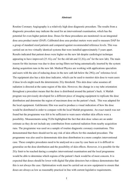
AbstractRoutine Coronary Angiography is a relatively high dose diagnostic procedure. The results from adiagnostic procedure may indicate the need for an interventional examination, which has thepotential for even higher patient dose. Doses for these procedures are monitored via an integrateddose area product meter (DAP). Calibrated dose area product meters were used to measure DAP fora group <strong>of</strong> standard sized patients and compared against recommended reference levels. This wascarried out on two virtually identical systems that were installed approximately 5 years apart.Results indicated that patient doses were higher on the new lab despite cardiologist techniqueappearing to have improved (25.1Gy.cm 2 for the old lab and 32.2Gy.cm 2 for the new lab). The mainreason for this increase was due to dose saving filters not being automatically inserted by the systemduring acquisition runs in the new lab. Medical Physics are working with applications specialistsand users with the aim <strong>of</strong> reducing doses in the new cath lab below the 29Gy.cm 2 reference level.The equipment also has a skin dose indicator, which can be used to monitor skin dose to warn usersif dose levels might reach the deterministic 2Gy threshold. This skin dose value assumes allradiation is directed at the same region <strong>of</strong> the skin. However, the change in x-ray tube orientationthroughout a procedure means that the dose is distributed around the patient’s back. A Matlabprogram was previously developed for a different piece <strong>of</strong> imaging equipment to replicate the dosedistribution and determine the region <strong>of</strong> maximum dose on the patient’s back. This was adapted forthe local equipment. Gafchromic film was used to produce a visual indication <strong>of</strong> how the dosewould be distributed in order to compare with the local Matlab programme. An exact match was notfound but the programme was felt to be sufficient to warn users whether skin effects were apossibility. Measurements using TLDs highlighted the fact that skin dose values are an underestimate as they do not include any contribution from scattered radiation from nearby acquisitionruns. The programme was used on a sample <strong>of</strong> routine diagnostic coronary examinations. Thisdemonstrated that there should not be any risk <strong>of</strong> skin effects for this standard procedure. Theprogramme was also used to demonstrate skin dose distribution in a more complex interventionalcase. These complex procedures need to be analysed on a case by case basis as it is difficult togeneralise on the dose distribution and the possibility <strong>of</strong> skin effects. However, it is possible for the2Gy limit to be reached during a complex interventional examination and the local programmewould be able to determine which regions <strong>of</strong> the patient’s back would be <strong>of</strong> most concern. It isexpected that doses should be lower with digital flat plate detectors but evidence demonstrates thatthis is not always the case. Optimisation work must be carried out on new equipment to ensure thatdoses are always as low as reasonably practical in line with current legislative requirements.I


