

Ghrelin's second life - World Journal of Gastroenterology
Ghrelin's second life - World Journal of Gastroenterology
Ghrelin's second life - World Journal of Gastroenterology

You also want an ePaper? Increase the reach of your titles
YUMPU automatically turns print PDFs into web optimized ePapers that Google loves.
A<br />
MCP-1 expression (fold change)<br />
8<br />
7<br />
6<br />
5<br />
4<br />
3<br />
2<br />
1<br />
0<br />
Control<br />
Protein<br />
Gene<br />
CCl4-7 wk<br />
CCl4-13 wk<br />
marrow to differentiate into hepatocytes. To assess the<br />
possibility <strong>of</strong> YGJ stimulating bone marrow cells to<br />
differentiate into hepatocytes, we investigated the presence<br />
<strong>of</strong> EGFP + /Alb + cells by double staining. In the<br />
CCl4injected control mice, the number <strong>of</strong> EGFP + cells<br />
increased steadily over time and the cells were distributed<br />
mainly in mesenchymal areas, but Alb + cells were<br />
mainly distributed in parenchymal areas, and there was no<br />
WJG|www.wjgnet.com<br />
CCl4-YGJ<br />
B 1 2 3 4<br />
C<br />
Wang XL et al . YGJ decoction protects against hepatic injury<br />
MCP-1<br />
GAPDH<br />
CCR2 expression (fold change)<br />
CCR2<br />
6<br />
5<br />
4<br />
3<br />
2<br />
1<br />
0<br />
Control<br />
Protein<br />
Gene<br />
b<br />
a<br />
CCl4-7 wk<br />
a<br />
b<br />
a<br />
c<br />
c<br />
26 KD<br />
42 KD<br />
36 KD<br />
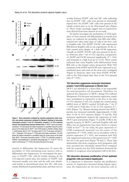
Figure 7 Gene expression analyzed by real-time polymerase chain reaction<br />
and protein expression analyzed by Western blotting <strong>of</strong> monocyte<br />
chemotaxis protein-1 and CC chemokine receptor 2 in CCl4-induced<br />
chronic liver injured bone marrow chimera mice. A: MCP-1 gene and protein<br />
expression. For both genes the relative expression is quantified using the control<br />
group as the basal level; B: MCP-1 and CCR2 bands by Western blotting. Band<br />
1, control group; band 2, CCl4 7 wk group; band 3, CCl4 13 wk group; and band 4,<br />
CCl4 + Yiguanjian (YGJ) group; C: CCR2 gene and protein expression. For both<br />
genes the relative expression is quantified using the control group as the basal<br />
level. MCP-1: Monocyte chemotaxis protein-1; CCR2: CC chemokine receptor 2;<br />
GAPDH: Glyceraldehydes-3- phosphate dehydrogenase. a P < 0.05, b P < 0.01 vs<br />
week 0 control group; c P < 0.05 vs the same time-point CCl4 group.<br />
b<br />
a<br />
CCl4-13 wk<br />
c<br />
CCl4-YGJ<br />
overlap between EGFP + cells and Alb + cells, indicating<br />
that no EGFP + /Alb + cells were present in chronically<br />
injured liver. No EGFP + /Alb + cells were present in the<br />
vehicle control mice or in the YGJtreated mice (Figure<br />
5). These results seemingly suggest that no hepatocytes<br />
were derived from bone marrow in our study.<br />
To further investigate the mechanisms <strong>of</strong> YGJ mediation<br />
<strong>of</strong> bone marrow cell differentiation in chronic liver<br />
injury, we explored the possibility that BM cells differentiated<br />
into other main types <strong>of</strong> cells in the liver, such<br />
as Kupffer cells. The EGFP + /F4/80 + cells represented<br />
BMderived Kupffer cells in our experiments. In the vehicle<br />
control mice, despite <strong>of</strong> a little F4/80 expression,<br />
virtually no EGFP + /F4/80 + cells were present in the liver.<br />
However, after 7 wk <strong>of</strong> CCl4 injection, coexpression<br />
<strong>of</strong> EGFP and F4/80 increased significantly (P < 0.01)<br />
and remained at a high level up to 13 wk. These results<br />
indicated that some Kupffer cells differentiated from<br />
BM cells as the hepatic injury progressed. With YGJ<br />
treatment, both EGFP and F4/80 were expressed in parenchymal<br />
areas, and some <strong>of</strong> the areas were overlapped<br />
(Figure 6). However, there were fewer EGFP + /F4/80 +<br />
cells in the YGJ-treated mice than in the CCl4-injected<br />
mice (P < 0.01).<br />
YGJ decoction suppresses monocyte chemotaxis<br />
protein-1 and CCR2 expression in fibrotic liver<br />
MCP1 was identified as a factor likely to be responsible<br />
for stem/progenitor cell recruitment. Therefore, we<br />
analyzed the expression <strong>of</strong> MCP1 during CCl4-induced<br />
fibrogenesis. For the gene and protein expression, results<br />
were normalized to expression in the liver at the time<br />
<strong>of</strong> CCl4 injection (0 wk), for example the control group<br />
mRNA level <strong>of</strong> MCP1 reached 2.8fold after 7 wk (P<br />
< 0.05) and 6.7fold after 13 wk <strong>of</strong> CCl4 injection (P <<br />
0.05) (Figure 7A). Western blotting showed that MCP1<br />
protein was increased markedly (approximately 4 fold)<br />
after 13 wk <strong>of</strong> CCl4 injection (Figure 7A and B). YGJ<br />
treatment significantly reduced the expression <strong>of</strong> both<br />
MCP-1 gene and protein (Figure 7A and B). CCR2 is the<br />
specific receptor <strong>of</strong> MCP1, and both Kupffer cells and<br />
HSCs, but not hepatocytes, have been shown to express<br />
CCR2 in liver [16] . In our study, the gene expression <strong>of</strong><br />
CCR2 continuously increased over the course <strong>of</strong> the<br />
experiment (P < 0.05), which was in accordance with its<br />
protein expression (P < 0.01), and treatment with YGJ<br />
only suppressed expression at the protein level (P < 0.05)<br />
but not at the gene level (Figure 7B and C). These data<br />
demonstrate that more exogenous stem/progenitor cells<br />
were recruited into chronically injured livers compared<br />
with the controls, but that YGJ treatment decreased the<br />
number <strong>of</strong> exogenous stem/progenitor cells in the liver.<br />
YGJ decoction decreases pro<strong>life</strong>ration <strong>of</strong> liver epithelial<br />
progenitor cells and mature hepatocytes<br />
It is important to determine whether the pro<strong>life</strong>rative<br />
activity <strong>of</strong> hepatocytes increased during hepatic injury<br />
since the hepatocytes are the most numerous cells in<br />
3244 July 7, 2012|Volume 18|Issue 25|


