NOVA Lite Skin Antibody 708330 - inova
NOVA Lite Skin Antibody 708330 - inova
NOVA Lite Skin Antibody 708330 - inova
You also want an ePaper? Increase the reach of your titles
YUMPU automatically turns print PDFs into web optimized ePapers that Google loves.
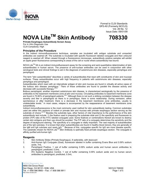
5. PBS Concentrate (40x), sufficient for 1000 mL6. Mounting Medium, 0.09% sodium azide, 7mL7. CoverslipsMaterials provided10 8-well <strong>Skin</strong> <strong>Antibody</strong> Substrate Slides1 7mL FITC Anti-Human IgG Conjugate1 0.5 mL Pemphigoid Positive1 0.5mL IFA Systems Negative Control1 25mL PBS Concentrate (40x)1 7mL Mounting Medium1 10 CoverslipsAdditional Materials Required But Not ProvidedMicropipets to deliver 15-1000μL volumeDistilled or deionized waterSqueeze bottles or Pasteur pipetsMoist chamber1L container (for diluting PBS)Coplin jarFluorescence microscope with 495nm exciter and 515nm barrier filterSpecimenThis procedure should be performed with a serum specimen. Addition of azide or other preservatives to the testsamples may adversely affect the results. Microbially contaminated, heat-treated, or specimens containing visibleparticulates should not be used. Grossly hemolyzed or lipemic serum specimens should be avoided.Following collection, the serum should be separated from the clot. NCCLS Document H18-A2 recommends thefollowing storage conditions for samples: 1) Store samples at room temperature no longer than 8 hours. 2) If theassay will not be completed within 8 hours, refrigerate the sample at 2 - 8 ° C. 3) If the assay will not be completedwithin 48 hrs, or for shipment of the sample, freeze at -20 ° C or lower. Frozen specimens must be mixed well afterthawing and prior to testing.Special Safety Precautions/Storage Conditions1. Store all the kit reagents at 2-8°C. Do not freeze. Reagents are stable until the expiration date when storedand handled as directed.2. Diluted PBS buffer is stable for 4 weeks at 2-8°CProcedural Notes:Warnings1. All human source material used in the preparation of controls for this product has been tested and foundnegative for antibody to HIV, HBsAg, and HCV by FDA cleared methods. No test method, however, canoffer complete assurance that HIV, HBV, HCV or other infectious agents are absent. Therefore, thePemphigoid Positive and IFA Systems Negative Control should be handled in the same manner aspotentially infectious material. 122. Sodium Azide is used as a preservative. Sodium Azide is a poison and may be toxic if ingested or absorbedthrough the skin or eyes. Sodium azide may react with lead or copper plumbing to form potentially explosivemetal azides. Flush sinks, if used for reagent disposal, with large volumes of water to prevent azide build-up.3. Use appropriate personal protective equipment while working with the reagents provided.4. Spilled reagents should be cleaned up immediately. Observe all federal, state and local environmentalregulations when disposing of wastes.Precautions1. This product is for In Vitro Diagnostic Use.2. Substitution of components other than those provided in this system may lead to inconsistent results.3. Incomplete or inefficient washing of IFA wells may cause high background.4. Adaptation of this assay for use with automated sample processors and other liquid handling devices, inwhole or in part, may yield differences in test results from those obtained using the manual procedure. It isthe responsibility of each laboratory to validate that their automated procedure yields test results withinacceptable limits.NCCLS <strong>708330</strong>Page 2 of 5
5. A variety of factors influence the assay performance. These include the starting temperature of the reagents,the strength of the microscope bulb used, the accuracy and reproducibility of the pipetting technique, thethoroughness of washing and the length of the incubation times during the assay. Careful attention toconsistency is required to obtain accurate and reproducible results.6. Over time, the Anti-Human IgG Conjugate may change in color due to exposure to light. However, the colorchange does not affect the assay performance.7. Strict adherence to the protocol is recommended.Quality ControlPemphigoid Positive and IFA System Negative Control should be run on every slide to insure that all reagents andprocedures perform properly. Additional suitable control sera may be prepared by aliquoting pooled human serumspecimens and storing at < -70 o C. In order for the test results to be considered valid, all of the criteria listed belowmust be met. If any of these are not met, the test results should be considered invalid and the assay repeated.1. The Pemphigoid Positive must be > 3+.2. The IFA System Negative Control must be negative.Procedure:Method Before you start1. Bring all reagents and samples to room temperature (20 - 26°C) and mix well.2. Dilute PBS Concentrate: IMPORTANT: Dilute the PBS Concentrate 1:40 by adding the contents of thePBS Concentrate bottle to 975mL of distilled or deionized water and mix thoroughly. The PBS buffer is usedfor diluting patient samples and as a wash buffer. The diluted buffer can be stored for up to 4 weeks at 2 -8°C.3. Dilute Patient Samples:a. Initial Screening: Dilute patient samples 1:10 with diluted PBS buffer (i.e., add 100μL of serum to900μL of PBS buffer).b. Titration: Make serial 2-fold dilutions from the initial screening dilution for all positive samples withdiluted PBS buffer (i.e. 1:20, 1:40,... 1: 2560).Assay Procedure1. Prepare Substrate Slides: Allow the substrate slide to reach room temperature prior to removal from itspouch. Label it with pencil and place it in a suitable moist chamber. Add 1 drop (70-90μL) of the undilutedpositive and the negative control to wells 1 and 2 respectively. Add 1 drop (70-90μL) of diluted patientsample to the remaining wells.2. Slide Incubation: Incubate the slide for 30 + 5 minutes in a moist chamber (a dampened paper towelplaced flat on the bottom of a closed plastic or glass container) will maintain the proper humidity conditions.Do not allow the substrate to dry out during the assay procedure.3. Wash Slides: After incubation, use a plastic squeeze bottle or pipet to gently wash off the serum withdiluted PBS buffer. Orient the slide and stream of PBS buffer so as to minimize wash-over of samplesbetween wells. Avoid directing the stream directly onto the wells to prevent substrate damage. Ifdesired, place the slides in a Coplin jar of diluted PBS buffer for up to 5 minutes.4. Addition of Fluorescent Conjugate: Shake off the excess PBS buffer. Place the slide back in the moistchamber and immediately cover each well with a drop of fluorescent conjugate. Incubate the slides for anadditional 30 + 5minutes.5. Wash Slides: Repeat Step 3.6. Coverslip: Coverslip procedures vary from lab to lab; however, the following procedure is recommended:a. Place a coverslip on a paper towel.b. Apply mounting medium in a continuous line to the bottom edge of the coverslip.c. Shake off the excess PBS buffer and touch the lower edge of the slide to the edge of the coverslip.Gently lower the slide onto the coverslip in such a way that the mounting medium flows to the topedge of the slide without air bubble formation or entrapment.Reporting Results:Interpretation of ResultsNegative Reaction. A sample is considered negative if specific staining of the intercellular areas, basementmembrane zone or other structures is equal to or less than the negative control. Samples can exhibit various degreesof background staining due to heterophile antibodies, or other specific reactions due to reticulin antibodies,anti-nuclear, anti-endomysial or anti-smooth muscle antibodies.Positive reaction for intercellular and basement membrane zone antibodies. A sample may be consideredpositive if specific staining of intercellular or basement membrane zone areas of the esophageal mucosa areobserved.NCCLS <strong>708330</strong>Page 3 of 5
Determine the fluorescence grade or intensity using these criteria:4+ Brilliant apple green fluorescence3+ Bright apple green fluorescence2+ Clearly distinguishable positive fluorescence1+ Lowest specific fluorescence that enables the intercellular or basement membrane zone areasstaining to be clearly differentiated from the background fluorescenceCalculationsN/AExpected Values & Specific Performance Characteristics<strong>NOVA</strong> <strong>Lite</strong> TM <strong>Skin</strong> <strong>Antibody</strong> substrate was used to test pemphigus and pemphigoid skin disease patients as well as100 random blood donors. The results appear below:Number PositivePatient Group Number Intercellular Basement Membrane ZonePemphigus 24 20 0Pemphigoid 18 0 12Normals 100 0 0Limitations of the Procedure1. The presence of skin autoantibodies is suggestive of certain autoimmune bullous skin diseases but shouldnot be considered diagnostic. The autoantibody result should be considered in combination with otherserological or biopsy results as well as the overall clinical history of the patient.2. In addition to skin autoantibodies, other autoantibodies can react with the esophagus substrate. Some ofthese include anti-nuclear (ANA), anti-mitochondrial (AMA), anti-smooth and anti-skeletal muscleautoantibodies. Presence of these antibodies should be noted and their presence should be confirmed andtitrated on other approved substrates.3. It should be noted that as a sequela to tissue damage, some burn patients produce weakly reactiveantibodies that also bind to intercellular areas. These so-called "pemphigus-like" antibodies give a weak anduneven staining of the mucosa, are transient in nature, and do not react with patient skin in vivo. 13,14,154. Because esophageal mucosa can contain certain blood group substances, anti-A and anti-B blood groupisoantibodies can react with esophagus. 16 The pattern of reactivity can, in some patients, mimic the patternobserved with pemphigus intercellular autoantibodies (although blood group antibody will also stain bloodcapillaries in the musculature of the esophagus section). Reactivity due to blood group antibody can beabsorbed out with blood group antigen. 175. A variety of external factors influence the test sensitivity including the type of fluorescence microscope used,the bulb strength and age, the magnification used, the filter system and the observer.6. If a band pass filter is used instead of a 515 barrier filter, increased artifactual staining may be observed.7. Only pencil should be used to label the slides. Use of any other writing material may cause artifactualstaining.8. All coplin jars used for slide washing should be free from all dye residues. Use of coplin jars containing dyeresidue may cause artifactual staining.9. Results of this assay should be used in conjunction with clinical findings and other serological tests.10. The assay performance characteristics have not been established for matrices other than serum.NCCLS <strong>708330</strong>Page 4 of 5
References1. Chorzelski TP, Beutner EH, Jablonska S: "The Role and Nature of Autoimmunity of Pemphigus" inImmunopathology of the <strong>Skin</strong>, second edition (EH Beutner, TP Chorzelski, SF Bean, eds.) John Wiley andSons, NY, pp. 183-229, 1979.2. Chorzelski TP, von Weiss JF, Lever WF: Clinical significance of autoantibodies in pemphigus. Arch.Dermatol.: 93:570, 1966.3. Jablonska S, et.al.: Pathogenesis of pemphigus erythematosus. Arch. Dermatol. Res. 258:135, 1977.4. Peck SM, et.al.: Studies in Bullous diseases. Arch. Dermatol. 103:141, 1971.5. Jordon RE, et.al.: Basement zone antibodies in bullous pemphigoid. JAMA 200:751, 1967.6. Person JR, Rogers RS: Bullous and cicatricial pemphigoid. Mayo Clin. Proc. 52:54,1977.7. Chorzelski TP, Jablonska S, Beutner EH: "Pemphigoid" in Immunopathology of the <strong>Skin</strong>, second edition (EHBeutner, TP Chorzelski, SF Bean, eds.) John Wiley and Sons, NY, pp. 243-255, 1979.8. Sabolinski ML, et.al.: Substrate specificity of anti-ephithelial antibodies of pemphigus vulgaris andpemphigus foliaceous sera in immunofluorescence tests on monkey and guinea pig esophagus sections. J.Invest. Dermatol. 88:545, 1987.9. O'Loughlin S, Goldman GC, Provost TT: Fate of pemphigus antibody following successful therapy.Preliminary evaluation of pemphigus antibody determination to regulate therapy. Arch. Dermatol. 114:1769,1978.10. Feibeleman C, Stolzner G, Provost TT: Pemphigus vulgaris. Superiority of monkey esophagus in thedetermination of pemphigus antibody. Arch. Dermatol. 117:561, 1981.11. Wiik A: Antinuclear factors in sera from healthy blood donors. Acta. Path. Microbiol. Scand. 84:215-220,1976.12. Biosafety in Microbiological and Biomedical Laboratories. Centers for Disease Control and Prevention/NationalInstitutes of Health, Fifth Edition, 200713. Ablin RJ, et.al.: Pemphigus-like antibodies in patients with skinburns. Vox Sang 16:73, 1969.14. Quismorio FP, Bland SL, Friou GJ: Autoimmunity in thermal injury: Occurrence of rheumatoid factors,antinuclear antibodies and antiepithelial antibodies. Clin. Exp. Immunol. 8:701, 1971.15. Trifthauser C, et.al.: Fluorescent antibody studies of pemphigus-like antibodies in burn sera. Ann. NY Acad.Sci. 177:227, 1971.16. Szulman AE: The histological distribution of blood group substances A and B in man. J. Exp. Medicine115:977, 1962.17. Beutner EH, et. al.: "Defined immunofluorescence-Basic concepts and their application to clinicalimmunodermatology." Immunopathology of the <strong>Skin</strong>, second edition (EH Beutner, TP Chorzelski, SF Bean,eds.) John Wiley and Sons, NY, pp. 29-75, 1979.I<strong>NOVA</strong> Diagnostics, Inc. 858-586-99009900 Old Grove Road 628330 Rev. 16San Diego, CA 921312009/AUGNCCLS <strong>708330</strong>Page 5 of 5


