


Create successful ePaper yourself
Turn your PDF publications into a flip-book with our unique Google optimized e-Paper software.
110 Chapter 14<br />
Choroid<br />
Choriocapillaris<br />
PEL<br />
Tight junction<br />
(blood–retina barrier)<br />
LRC<br />
Cone<br />
Rod<br />
OLM<br />
Visual cortex (area 17)<br />
ONL<br />
Optic<br />
nerve<br />
Pedicle<br />
OPL<br />
Lateral geniculate body<br />
Optic tract INL<br />
Optic chiasm<br />
Oligodendrocytes<br />
IPL<br />
Sphericle<br />
Capillary plexus<br />
Horizontal cell<br />
Bipolar cell<br />
Amacrine cell<br />
Müller cell<br />
Optic disk<br />
Scleral canal<br />
NFL<br />
GCL<br />
Ganglion cell<br />
Basement<br />
membrane<br />
ILM<br />
Vitreous body<br />
Central<br />
artery of<br />
retina<br />
Light<br />
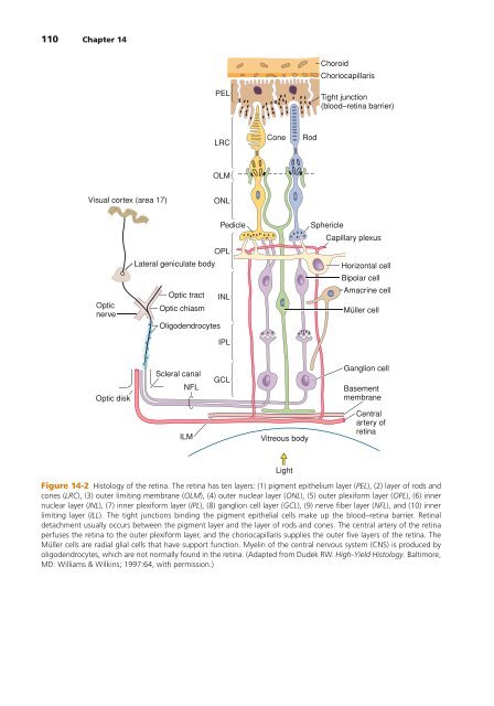
Figure 14-2 Histology of the retina. The retina has ten layers: (1) pigment epithelium layer (PEL), (2) layer of rods and<br />
cones (LRC), (3) outer limiting membrane (OLM), (4) outer nuclear layer (ONL), (5) outer plexiform layer (OPL), (6) inner<br />
nuclear layer (INL), (7) inner plexiform layer (IPL), (8) ganglion cell layer (GCL), (9) nerve fiber layer (NFL), and (10) inner<br />
limiting layer (ILL). The tight junctions binding the pigment epithelial cells make up the blood–retina barrier. Retinal<br />
detachment usually occurs between the pigment layer and the layer of rods and cones. The central artery of the retina<br />
perfuses the retina to the outer plexiform layer, and the choriocapillaris supplies the outer five layers of the retina. The<br />
Müller cells are radial glial cells that have support function. Myelin of the central nervous system (CNS) is produced by<br />
oligodendrocytes, which are not normally found in the retina. (Adapted from Dudek RW. <strong>High</strong>-<strong>Yield</strong> Histology. Baltimore,<br />
MD: Williams & Wilkins; 1997:64, with permission.)