


Create successful ePaper yourself
Turn your PDF publications into a flip-book with our unique Google optimized e-Paper software.
14 Chapter 2<br />
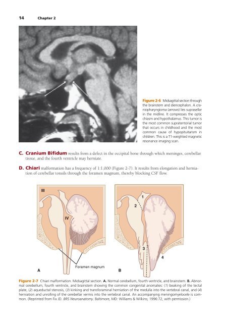
Figure 2-6 Midsagittal section through<br />
the brainstem and diencephalon. A craniopharyngioma<br />
(arrows) lies supra sellar<br />
in the midline. It compresses the optic<br />
chiasm and hypothalamus. This tumor is<br />
the most common supratentorial tumor<br />
that occurs in childhood and the most<br />
common cause of hypopituitarism in<br />
children. This is a T1-weighted magnetic<br />
resonance imaging scan.<br />
C. Cranium Bifidum results from a defect in the occipital bone through which meninges, cerebellar<br />
tissue, and the fourth ventricle may herniate.<br />
D. Chiari malformation has a frequency of 1:1,000 (Figure 2-7). It results from elongation and herniation<br />
of cerebellar tonsils through the foramen magnum, thereby blocking CSF flow.<br />
III<br />
1<br />
2<br />
IV<br />
3<br />
4<br />
A<br />
Foramen magnum<br />
B<br />
Figure 2-7 Chiari malformation. Midsagittal section. A. Normal cerebellum, fourth ventricle, and brainstem. B. Abnormal<br />
cerebellum, fourth ventricle, and brainstem showing the common congenital anomalies: (1) beaking of the tectal<br />
plate, (2) aqueductal stenosis, (3) kinking and transforaminal herniation of the medulla into the vertebral canal, and (4)<br />
herniation and unrolling of the cerebellar vermis into the vertebral canal. An accompanying meningomyelocele is common.<br />
(Reprinted from Fix JD. BRS <strong>Neuroanatomy</strong>. Baltimore, MD: Williams & Wilkins; 1996:72, with permission.)