


Create successful ePaper yourself
Turn your PDF publications into a flip-book with our unique Google optimized e-Paper software.
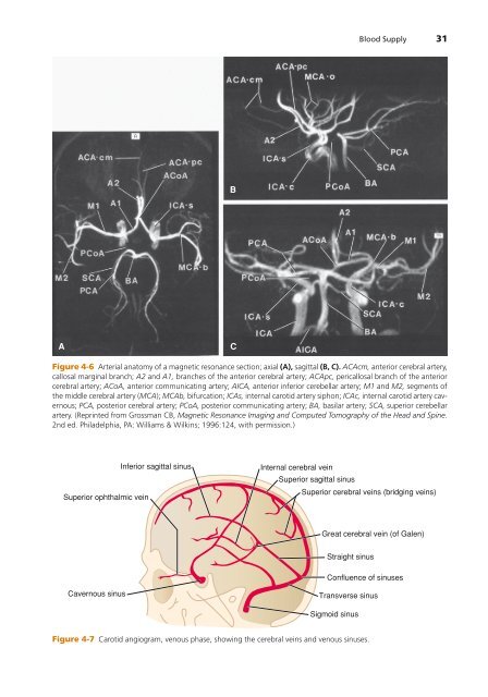
Blood Supply 31<br />
B<br />
A<br />
C<br />
Figure 4-6 Arterial anatomy of a magnetic resonance section; axial (A), sagittal (B, C). ACAcm, anterior cerebral artery,<br />
callosal marginal branch; A2 and A1, branches of the anterior cerebral artery; ACApc, pericallosal branch of the anterior<br />
cerebral artery; ACoA, anterior communicating artery; AICA, anterior inferior cerebellar artery; M1 and M2, segments of<br />
the middle cerebral artery (MCA); MCAb, bifurcation; ICAs, internal carotid artery siphon; ICAc, internal carotid artery cavernous;<br />
PCA, posterior cerebral artery; PCoA, posterior communicating artery; BA, basilar artery; SCA, superior cerebellar<br />
artery. (Reprinted from Grossman CB, Magnetic Resonance Imaging and Computed Tomography of the Head and Spine.<br />
2nd ed. Philadelphia, PA: Williams & Wilkins; 1996:124, with permission.)<br />
Superior ophthalmic vein<br />
Inferior sagittal sinus<br />
Internal cerebral vein<br />
Superior sagittal sinus<br />
Superior cerebral veins (bridging veins)<br />
Great cerebral vein (of Galen)<br />
Straight sinus<br />
Confluence of sinuses<br />
Cavernous sinus<br />
Transverse sinus<br />
Sigmoid sinus<br />
Figure 4-7 Carotid angiogram, venous phase, showing the cerebral veins and venous sinuses.