Dental Asia March/April 2023
For more than two decades, Dental Asia is the premium journal in linking dental innovators and manufacturers to its rightful audience. We devote ourselves in showcasing the latest dental technology and share evidence-based clinical philosophies to serve as an educational platform to dental professionals. Our combined portfolio of print and digital media also allows us to reach a wider market and secure our position as the leading dental media in the Asia Pacific region while facilitating global interactions among our readers.
For more than two decades, Dental Asia is the premium journal in linking dental innovators
and manufacturers to its rightful audience. We devote ourselves in showcasing the latest dental technology and share evidence-based clinical philosophies to serve as an educational platform to dental professionals. Our combined portfolio of print and digital media also allows us to reach a wider market and secure our position as the leading dental media in the Asia Pacific region while facilitating global interactions among our readers.

You also want an ePaper? Increase the reach of your titles
YUMPU automatically turns print PDFs into web optimized ePapers that Google loves.
CLINICAL FEATURE<br />
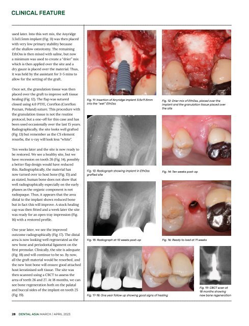
used later. Into this wet mix, the Anyridge<br />
3.5x11.5mm implant (Fig. 11) was then placed<br />
with very low primary stability because<br />
of the shallow osteotomy. The remaining<br />
EthOss is then mixed with saline, but now<br />
a minimum was used to create a “drier” mix<br />
which is then applied over the site and a<br />
dry gauze is placed over the material. Thus,<br />
it was held by the assistant for 3-5 mins to<br />
allow for the setting of the graft.<br />
Once set, the granulation tissue was then<br />
placed over the graft to improve soft tissue<br />
healing (Fig. 12). The flap was sutured<br />
closed using 4.0 PTFE, Coreflon (Coreflon<br />
Poznan, Poland) suture. This procedure with<br />
the granulation tissue is not the routine<br />
protocol, but a one-off for this case and has<br />
been used occasionally over the last 15 years.<br />
Radiographically, the site looks well grafted<br />
(Fig. 13) but remember as the CS element<br />
resorbs, the x-ray will look less “white”.<br />
Fig. 11: Insertion of Anyridge implant 3.5x11.5mm<br />
into the “wet” EthOss<br />
Fig. 12: Drier mix of EthOss, placed over the<br />
implant and the granulation tissue placed over<br />
the site<br />
Ten weeks later and the site is now ready to<br />
be restored. We see a healthy site, but we<br />
have recession on tooth 26 (Fig. 14), possibly<br />
a better flap design would have reduced<br />
this. Radiographically, the material has<br />
now turned over to host bone (Fig. 15) and<br />
as stated, human bone does not show that<br />
well radiographically especially on the early<br />
phases as the organic component is not<br />
radiopaque. Thus, it appears that the area<br />
distal to the implant shows reduced bone<br />
but in fact this will improve. A stock healing<br />
cap was then fitted and a week later the site<br />
was ready for an open tray impression (Fig.<br />
16) with a restored profile.<br />
Fig. 13: Radiograph showing implant in EthOss<br />
grafted site<br />
Fig. 14: Ten weeks post-op<br />
One year later, we see the improved<br />
outcome radiographically (Fig. 17). The distal<br />
area is now looking well regenerated as the<br />
new bone and periodontal ligament on the<br />
first premolar. Clinically, the site is adequate<br />
(Fig. 18) and will continue to be so. By now,<br />
all the graft material would be resorbed, and<br />
the new host bone will ensure good attached<br />
host keratinised soft tissue. The site was<br />
then scanned using a CBCT to assess the<br />
area of teeth 26 and 27. At 18 months, we can<br />
see bone regeneration both on the palatal<br />
and buccal sides of the implant on tooth 25<br />
(Fig. 19).<br />
Fig. 15: Radiograph at 10 weeks post-op<br />
Fig. 17-18: One year follow up showing good signs of healing<br />
Fig. 16: Ready to load at 11 weeks<br />
Fig. 19: CBCT scan at<br />
18 months showing<br />
new bone regeneration<br />
28 DENTAL ASIA MARCH / APRIL <strong>2023</strong>