Dental Asia March/April 2023
For more than two decades, Dental Asia is the premium journal in linking dental innovators and manufacturers to its rightful audience. We devote ourselves in showcasing the latest dental technology and share evidence-based clinical philosophies to serve as an educational platform to dental professionals. Our combined portfolio of print and digital media also allows us to reach a wider market and secure our position as the leading dental media in the Asia Pacific region while facilitating global interactions among our readers.
For more than two decades, Dental Asia is the premium journal in linking dental innovators
and manufacturers to its rightful audience. We devote ourselves in showcasing the latest dental technology and share evidence-based clinical philosophies to serve as an educational platform to dental professionals. Our combined portfolio of print and digital media also allows us to reach a wider market and secure our position as the leading dental media in the Asia Pacific region while facilitating global interactions among our readers.
Create successful ePaper yourself
Turn your PDF publications into a flip-book with our unique Google optimized e-Paper software.
CLINICAL FEATURE<br />
• Pecking motion, light touch, stable finger<br />
rests<br />
• Wipe the instrument clean of debris after<br />
use in a canal<br />
• Examine flutes and the file tip, preferably<br />
under magnification.<br />
• Copious irrigation for flushing debris<br />
between instruments and the use of rotary<br />
instruments with an irrigant filled canal<br />
• Discard files regularly, especially after use<br />
in calcified, narrow or curved canals.<br />
• Familiarity and hands-on practice with the<br />
instrumentation technique<br />
Teeth presenting with concomitant periapical/<br />
periradicular lesions and a fractured<br />
instrument in the apical third are deemed<br />
complex endodontic cases. When a patient<br />
presents with a separated instrument, the<br />
advantages and disadvantages of attempting<br />
instrument retrieval should always be weighed.<br />
The clinician’s skillset, clinical experience and<br />
armamentarium available at his disposal play<br />
an important part in conservative instrument<br />
retrieval.<br />
Retrieval of the separated instruments with least<br />
removal of root dentin should be the highest<br />
priority. Critical prognostic determinants for<br />
instrument retrieval include the position of the<br />
fractured instrument, length of the separated<br />
instrument, canal curvature and visibility of<br />
the coronal end of the instrument. Moreover,<br />
canal anatomy, including the diameter, length<br />
and level of curvature of the canal, as well as<br />
the thickness of root dentin and the depth of<br />
external concavities will influence the safe<br />
removal of the separated instrument fragment.<br />
The dentinal engagement of the separated<br />
fragment at multiple sites cannot be efficiently<br />
overcome by ultrasonics alone, especially<br />
when the fragment length exceeds 3-4mm. A<br />
lasso tool or loop device is then necessary for<br />
predictable retrieval.<br />
Other relevant clinical findings for this case<br />
included heavy buttressing of alveolar bone<br />
including the entire upper anterior area,<br />
and an extremely deep anterior bite with<br />
retroclined incisors. The four upper incisors<br />
had a history of multiple repetitive nontraumatic<br />
fractures treated by endodontics<br />
and zirconia full coverage crowns pointing<br />
to the absence of a mutually protective<br />
occlusion. The patient had not received an<br />
orthodontic or restorative treatment for<br />
correction of his bite or occlusal overloads.<br />
In this scenario, the long-term impact on<br />
canines could account for localised trauma<br />
and a probable cause for cervical root<br />
resorption. The specific site of resorption,<br />
the disto-labial root surface at the cervical<br />
third could also correlate to this traumatic<br />
bite. The follow-up radiograph at six months<br />
showed complete healing but partial<br />
replacement or resorption of the repair<br />
material, MTA. This could be possibly caused<br />
by the persistence of excessive occlusal<br />
forces on the tooth, specific to the said area.<br />
The patient has not initiated any orthodontic<br />
treatment and is reluctant to do so. An<br />
occlusal appliance like a splint could be<br />
protective and thus useful in preventing or<br />
minimising diurnal non-functional forces.<br />
A single, localised and accessible site of<br />
EICRR allowed for surgical direct access and<br />
management. This allowed for immediate<br />
intervention as well as predictability in<br />
terms of direct visualisation and treatment.<br />
MTA is a bioactive, sterile, radiopaque and<br />
dimensionally stable material not sensitive<br />
to moisture and blood contamination. MTA<br />
provides an effective seal against dentin and<br />
cementum and promotes biologic repair and<br />
regeneration of the periodontal ligament and<br />
bone, cementum and dentin. Other alternate<br />
bioactive materials like bioceramics may also<br />
be used.<br />
Alternate treatment modalities for EICRR<br />
include long-term calcium hydroxide<br />
intracanal dressings alone or in conjunction<br />
with corticosteroids. Although they may<br />
attenuate the progression, they may not<br />
predictably arrest resorption. Regenerative<br />
endodontics and intentional replantation<br />
procedures have also been used for the<br />
management of EICRR.<br />
CONCLUSION<br />
Instrument retrieval and conservation of<br />
root dentin are critical for the long-term<br />
prognosis and predictable endodontic<br />
outcome. The present case had an added<br />
dimension of EICRR that was managed<br />
via a surgical approach and restored with<br />
a bioactive material, MTA. A six-month<br />
follow-up demonstrated adequate clinical<br />
and radiographic healing. The long-term<br />
prognosis of the tooth would ultimately<br />
depend on the removal or diminution of<br />
multiple etiologic factors. DA<br />
REFERENCES:<br />
Terauchi Y et al. Present status and future<br />
directions: Removal of fractured instruments. Int<br />
Endod J. 2022 May; Suppl 3:685-709.<br />
Abbott PV. Prevention and management of<br />
external inflammatory resorption following trauma<br />
to teeth. Aust Dent J. 2016 Mar; Suppl 1:82-94.<br />
Saoud TM et al. Regeneration and Repair in<br />
Endodontics-A Special Issue of the Regenerative<br />
Endodontics-A New Era in Clinical Endodontics.<br />
Dent J (Basel). 2016; 27:4(1):3.<br />
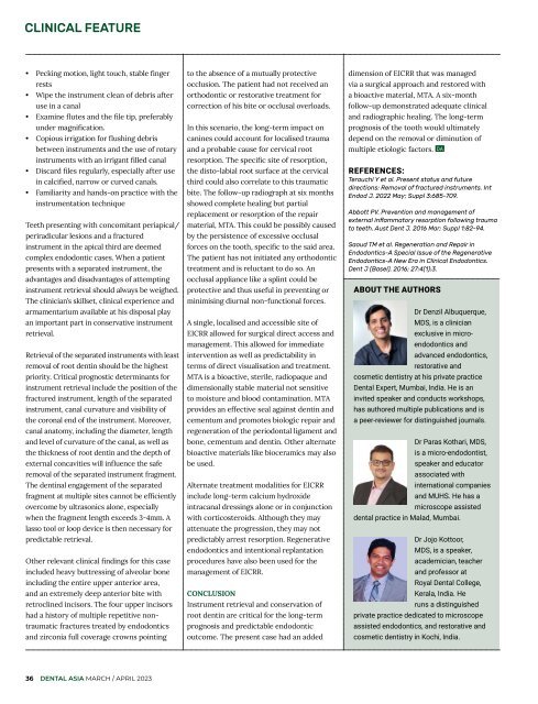
ABOUT THE AUTHORS<br />
Dr Denzil Albuquerque,<br />
MDS, is a clinician<br />
exclusive in microendodontics<br />
and<br />
advanced endodontics,<br />
restorative and<br />
cosmetic dentistry at his private practice<br />
<strong>Dental</strong> Expert, Mumbai, India. He is an<br />
invited speaker and conducts workshops,<br />
has authored multiple publications and is<br />
a peer-reviewer for distinguished journals.<br />
Dr Paras Kothari, MDS,<br />
is a micro-endodontist,<br />
speaker and educator<br />
associated with<br />
international companies<br />
and MUHS. He has a<br />
microscope assisted<br />
dental practice in Malad, Mumbai.<br />
Dr Jojo Kottoor,<br />
MDS, is a speaker,<br />
academician, teacher<br />
and professor at<br />
Royal <strong>Dental</strong> College,<br />
Kerala, India. He<br />
runs a distinguished<br />
private practice dedicated to microscope<br />
assisted endodontics, and restorative and<br />
cosmetic dentistry in Kochi, India.<br />
36 DENTAL ASIA MARCH / APRIL <strong>2023</strong>


