

Dental Asia November/December 2023
For more than two decades, Dental Asia is the premium journal in linking dental innovators and manufacturers to its rightful audience. We devote ourselves in showcasing the latest dental technology and share evidence-based clinical philosophies to serve as an educational platform to dental professionals. Our combined portfolio of print and digital media also allows us to reach a wider market and secure our position as the leading dental media in the Asia Pacific region while facilitating global interactions among our readers.
For more than two decades, Dental Asia is the premium journal in linking dental innovators and manufacturers to its rightful audience. We devote ourselves in showcasing the latest dental technology and share evidence-based clinical philosophies to serve as an educational platform to dental professionals. Our combined portfolio of print and digital media also allows us to reach a wider market and secure our position as the leading dental media in the Asia Pacific region while facilitating global interactions among our readers.

You also want an ePaper? Increase the reach of your titles
YUMPU automatically turns print PDFs into web optimized ePapers that Google loves.
CLINICAL FEATURE<br />
3D-printer technique for<br />
restoration of secondclass<br />
cavities in molars<br />
without conventional septal<br />
techniques: Case report<br />
By Dr Nokame Yoshinori<br />
The use of a septum is conventionally<br />
required for the restoration of secondclass<br />
cavities in molars.<br />
However, a problem with the septal<br />
technique is the difficulty in obtaining<br />
a perfect fit at the microscopic level. If<br />
there is even a small gap, the restoration<br />
will leak and harden, creating a bur<br />
that is difficult to correct. Although it is<br />
possible to create a single abutment, it is<br />
difficult to create an abutment consisting<br />
of multiple abutments like a natural<br />
tooth.<br />
Especially in the case of maxillary first<br />
premolars, the second-class cavity on the<br />
proximal side is difficult to create.<br />
The reason is that maxillary first<br />
premolars are narrowed proximally<br />
down to the root of the tooth,<br />
which often cause concavity at the<br />
base of the fossa, making it difficult<br />
to fit a septal wall. The crowns<br />
often have two abutments on the<br />
proximal adjacent surfaces. This<br />
has a functional meaning in that<br />
the buccal marginal ridge is aligned<br />
with the smaller cuspid to form an<br />
aesthetic and easy-to-clean drumshaped<br />
void.<br />
Therefore, to restore the proximal<br />
second-level socket of the maxillary<br />
first premolar to its original natural<br />
tooth form with composite resin, new<br />
restorative methods other than septal<br />
technique are needed.<br />
CASE REPORT<br />
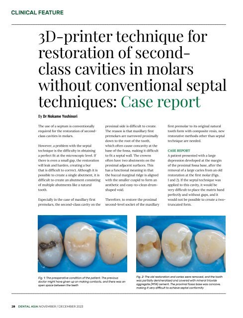
A patient presented with a large<br />
depression developed at the margin<br />
of the proximal fossa base, after the<br />
removal of a large caries from an old<br />
restoration at the first molar (Figs.<br />
1 and 2). If the septal technique was<br />
applied to this cavity, it would be<br />
very difficult to place the matrix band<br />
perfectly and without gaps, and it<br />
would not be possible to create a twotruncated<br />
form.<br />
Fig. 1: The preoperative condition of the patient. The previous<br />
doctor might have given up on making contacts, and there was an<br />
open space between the teeth<br />
Fig. 2: The old restoration and caries were removed, and the tooth<br />
was partially demineralised and covered with mineral trioxide<br />
aggregate (MTA) cement. The proximal fossa base was concave,<br />
making it very difficult to achieve septal conformity<br />
28 DENTAL ASIA NOVEMBER / DECEMBER <strong>2023</strong>


