

Dental Asia November/December 2023
For more than two decades, Dental Asia is the premium journal in linking dental innovators and manufacturers to its rightful audience. We devote ourselves in showcasing the latest dental technology and share evidence-based clinical philosophies to serve as an educational platform to dental professionals. Our combined portfolio of print and digital media also allows us to reach a wider market and secure our position as the leading dental media in the Asia Pacific region while facilitating global interactions among our readers.
For more than two decades, Dental Asia is the premium journal in linking dental innovators and manufacturers to its rightful audience. We devote ourselves in showcasing the latest dental technology and share evidence-based clinical philosophies to serve as an educational platform to dental professionals. Our combined portfolio of print and digital media also allows us to reach a wider market and secure our position as the leading dental media in the Asia Pacific region while facilitating global interactions among our readers.

Create successful ePaper yourself
Turn your PDF publications into a flip-book with our unique Google optimized e-Paper software.
CLINICAL FEATURE<br />
Shofu Beautifil LS II low-shrinkage composite<br />
(0.85%). In this manner, a natural form and<br />
function was created (Fig. 4).<br />
To build tooth 46, the temporary cement was<br />
removed and a caries dye was applied with<br />
caries endpoint removal. The mesial floor was<br />
almost subgingival, so a deep margin elevation<br />
was decided using a palodent wedge to seal<br />
this critical area from any sulcus fluid coming<br />
from the dam (Fig. 5).<br />
The entire preparation was sandblasted with<br />
50µ aluminum oxide powder for 7-8secs.<br />
Air abrasion compacted the smear layer<br />
in peritubular dentine and thinned the<br />
intertubular dentine.<br />
This ensured the removal of any leftover<br />
temporary cement to provide a cleaner surface<br />
to achieve a predictable bonding strength<br />
over 40MPa. The weakest part of the tooth<br />
was noted, which informed the building of a<br />
biobase incorporating polyethylene fibres<br />
(Fig. 6). With this procedure, the structurally<br />
compromised tooth was protected from<br />
shrinkage stress which otherwise would<br />
cause catastrophic failure of the entire tooth<br />
structure.<br />
A 6th Gen Shofu FL-Bond II was applied on<br />
the tooth using the proper protocol according<br />
to the provided instruction manual. The<br />
6th Gen Shofu FL-Bond is a gold-standard<br />
bonding agent with a high level of bonding<br />
strength to both dentin and enamel. The<br />
bonding strength would not decrease in the<br />
future as compared to a one bottle system.<br />
Once the bond was cured, a thin layer of<br />
2nd Gen Shofu Injectable XSL (slow levelling)<br />
high-strength composite was applied over<br />
the tooth. Care should be taken to ensure a<br />
thickness no more than 0.5mm and curing<br />
should only be done after placement of<br />
Ribbond fibres.<br />
Next, 3mm Ribbond fibres were cut using a<br />
special Ribbond scissors into small pieces<br />
and dipped in unfilled, 4th Gen bond resin,<br />
drying the excess of the bonding agent.<br />
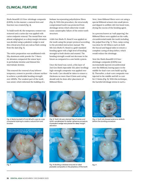
In a process known as ‘wall-papering’, the<br />
Ribbond fibres were applied on the walls,<br />
circumferential inside the tooth including<br />
the pulpal floor (Fig. 7). Then, ramp curing<br />
was done for 40-60secs each on both<br />
the buccal and lingual sides to ensure a<br />
complete curing of deep defect, which<br />
would reduce the shrinkage.<br />
Next the Shofu Beautifil LS II lowshrinkage<br />
composite (0.85%) was<br />
incrementally layered circumferential<br />
over the Ribbond, leaving space in the<br />
middle for dual-cure core build-up (Fig.<br />
8). Thereafter, a dual-cure composite was<br />
injected in the middle and left to cure<br />
for 2-3mins (Fig. 9). With this technique,<br />
the harmful shrinkage stress in such a<br />
Fig. 4: Restoring teeth 47 and 48 with cusp-by-cusp<br />
incremental layering to create a natural form and<br />
function<br />
Fig. 5: Tooth 46 was cleaned free of caries and<br />
tooth-sandblasted for better adhesive procedure.<br />
A palodent wedge was placed for deep margin<br />
elevation<br />
Fig. 6: Tooth 46 showed extensive defects<br />
before the bonding procedure<br />
Fig. 7 Fig. 8: Building a biobase ensured an ideal<br />
Fig. 9<br />
packable composite on the periphery of the tooth<br />
32 DENTAL ASIA NOVEMBER / DECEMBER <strong>2023</strong>


