Dental Asia November/December 2023
For more than two decades, Dental Asia is the premium journal in linking dental innovators and manufacturers to its rightful audience. We devote ourselves in showcasing the latest dental technology and share evidence-based clinical philosophies to serve as an educational platform to dental professionals. Our combined portfolio of print and digital media also allows us to reach a wider market and secure our position as the leading dental media in the Asia Pacific region while facilitating global interactions among our readers.
For more than two decades, Dental Asia is the premium journal in linking dental innovators and manufacturers to its rightful audience. We devote ourselves in showcasing the latest dental technology and share evidence-based clinical philosophies to serve as an educational platform to dental professionals. Our combined portfolio of print and digital media also allows us to reach a wider market and secure our position as the leading dental media in the Asia Pacific region while facilitating global interactions among our readers.

You also want an ePaper? Increase the reach of your titles
YUMPU automatically turns print PDFs into web optimized ePapers that Google loves.
CLINICAL FEATURE<br />
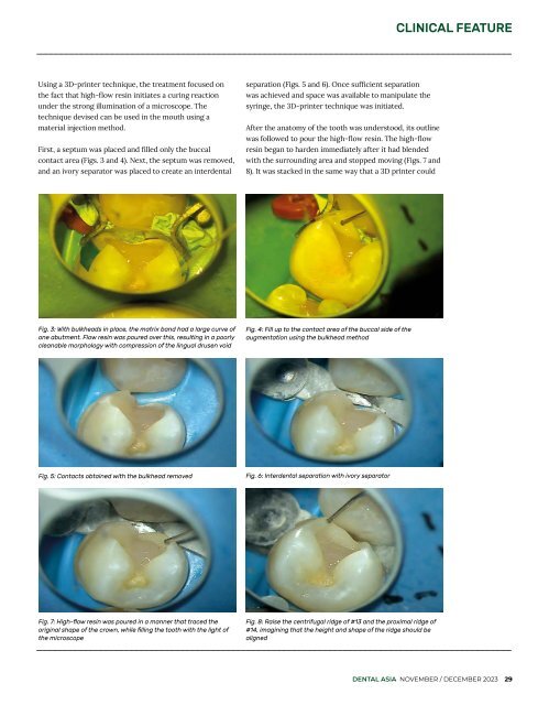
Using a 3D-printer technique, the treatment focused on<br />
the fact that high-flow resin initiates a curing reaction<br />
under the strong illumination of a microscope. The<br />
technique devised can be used in the mouth using a<br />
material injection method.<br />
First, a septum was placed and filled only the buccal<br />
contact area (Figs. 3 and 4). Next, the septum was removed,<br />
and an ivory separator was placed to create an interdental<br />
separation (Figs. 5 and 6). Once sufficient separation<br />
was achieved and space was available to manipulate the<br />
syringe, the 3D-printer technique was initiated.<br />
After the anatomy of the tooth was understood, its outline<br />
was followed to pour the high-flow resin. The high-flow<br />
resin began to harden immediately after it had blended<br />
with the surrounding area and stopped moving (Figs. 7 and<br />
8). It was stacked in the same way that a 3D printer could<br />
Fig. 3: With bulkheads in place, the matrix band had a large curve of<br />
one abutment. Flow resin was poured over this, resulting in a poorly<br />
cleanable morphology with compression of the lingual drusen void<br />
Fig. 4: Fill up to the contact area of the buccal side of the<br />
augmentation using the bulkhead method<br />
Fig. 5: Contacts obtained with the bulkhead removed<br />
Fig. 6: Interdental separation with ivory separator<br />
Fig. 7: High-flow resin was poured in a manner that traced the<br />
original shape of the crown, while filling the tooth with the light of<br />
the microscope<br />
Fig. 8: Raise the centrifugal ridge of #13 and the proximal ridge of<br />
#14, imagining that the height and shape of the ridge should be<br />
aligned<br />
DENTAL ASIA NOVEMBER / DECEMBER <strong>2023</strong> 29