Dental Asia November/December 2023
For more than two decades, Dental Asia is the premium journal in linking dental innovators and manufacturers to its rightful audience. We devote ourselves in showcasing the latest dental technology and share evidence-based clinical philosophies to serve as an educational platform to dental professionals. Our combined portfolio of print and digital media also allows us to reach a wider market and secure our position as the leading dental media in the Asia Pacific region while facilitating global interactions among our readers.
For more than two decades, Dental Asia is the premium journal in linking dental innovators and manufacturers to its rightful audience. We devote ourselves in showcasing the latest dental technology and share evidence-based clinical philosophies to serve as an educational platform to dental professionals. Our combined portfolio of print and digital media also allows us to reach a wider market and secure our position as the leading dental media in the Asia Pacific region while facilitating global interactions among our readers.

Create successful ePaper yourself
Turn your PDF publications into a flip-book with our unique Google optimized e-Paper software.
CLINICAL FEATURE<br />
Quadrant Dentistry: Managing<br />
structurally compromised root<br />
canal treated molar tooth using<br />
biomimetic principles, fibre<br />
reinforcement and adhesive<br />
treatment By Dr Anand Narvekar<br />
CASE HISTORY<br />
A 35-year-old female complained of pain<br />
during the consumption of hot and cold<br />
beverages for several months. Due to the<br />
COVID-19 pandemic, she delayed her visit<br />
to the clinic.<br />
Clinical examination revealed large carious<br />
two walls defect on tooth 46. IOPA showed<br />
the involvement of nerve with no signs of<br />
periapical infection and widening for PDL<br />
space. Tooth 47 showed marginal leakage<br />
from old composite restorations and tooth<br />
48 revealed a Class 1 defect (Figs. 1 and 2).<br />
TREATMENT PLAN<br />
Phase 1: Root canal treatment<br />
Caries-driven access for root canal<br />
treatment with complete removal of caries.<br />
Canal shaping was done with Endostar<br />
azure file system. 03 canal system with<br />
02 mesials and 01 wide distal. Root canal<br />
therapy was done in single visit.<br />
Phase 2: Quadrant direct restorations and<br />
overlay preparations<br />
The patient was recalled after 15 days for<br />
post-root canal build and direct restorations<br />
for teeth 47 and 48.<br />
Quadrant isolation was done from teeth 42-<br />
48, with the placement of a wingless active<br />
clamp on tooth 48. A heavy rubber dam was<br />
selected for a better inversion and perfect<br />
seal due to the structurally compromised<br />
tooth 46 (Fig. 3).<br />
When performing a quadrant treatment<br />
approach in one setting, it is recommended<br />
to start with the last tooth as the patient’s<br />
jaw would close as time elapses, which<br />
presents difficulty when working on the last<br />
posterior tooth 7 or 8.<br />
Old composite restoration was removed<br />
from tooth 47 and caries were removed from<br />
teeth 47 and 48, with the help of a caries dye.<br />
Incremental morphological-driven cuspby-cusp<br />
layering was selected to reduce the<br />
C-factor as Class 1 defects produce a high<br />
C-factor of 5, which leads to polymerisation<br />
shrinkage and shrinkage stress.<br />
The first layer was applied using Shofu<br />
Injectable XSL, a 2nd Gen high compressive<br />
strength composite. This layer must be<br />
minimally 0.5mm as it will seal the uneven<br />
dentine floor and protect the hybrid layer<br />
from the polymerisation shrinkage stress.<br />
The second layer was applied using the<br />
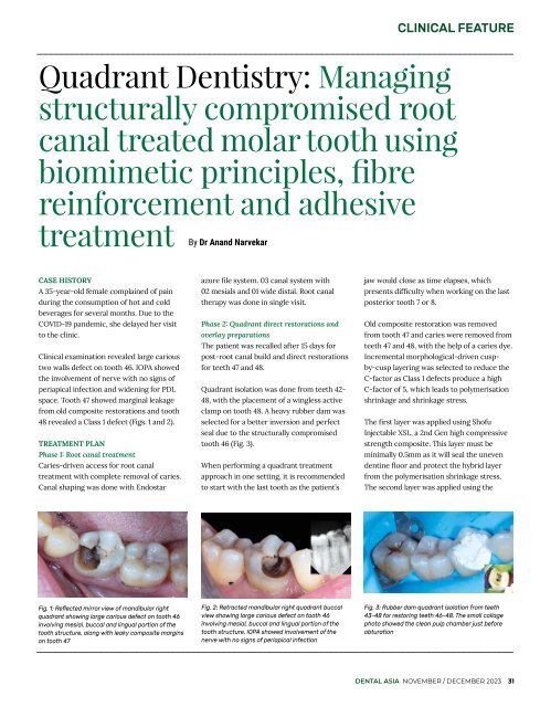
Fig. 1: Reflected mirror view of mandibular right<br />
quadrant showing large carious defect on tooth 46<br />
involving mesial, buccal and lingual portion of the<br />
tooth structure, along with leaky composite margins<br />
on tooth 47<br />
Fig. 2: Retracted mandibular right quadrant buccal<br />
view showing large carious defect on tooth 46<br />
involving mesial, buccal and lingual portion of the<br />
tooth structure. IOPA showed involvement of the<br />
nerve with no signs of periapical infection<br />
Fig. 3: Rubber dam quadrant isolation from teeth<br />
43-48 for restoring teeth 46-48. The small collage<br />
photo showed the clean pulp chamber just before<br />
obturation<br />
DENTAL ASIA NOVEMBER / DECEMBER <strong>2023</strong> 31