

Dental Asia January/February 2024
For more than two decades, Dental Asia is the premium journal in linking dental innovators and manufacturers to its rightful audience. We devote ourselves in showcasing the latest dental technology and share evidence-based clinical philosophies to serve as an educational platform to dental professionals. Our combined portfolio of print and digital media also allows us to reach a wider market and secure our position as the leading dental media in the Asia Pacific region while facilitating global interactions among our readers.
For more than two decades, Dental Asia is the premium journal in linking dental innovators and manufacturers to its rightful audience. We devote ourselves in showcasing the latest dental technology and share evidence-based clinical philosophies to serve as an educational platform to dental professionals. Our combined portfolio of print and digital media also allows us to reach a wider market and secure our position as the leading dental media in the Asia Pacific region while facilitating global interactions among our readers.

You also want an ePaper? Increase the reach of your titles
YUMPU automatically turns print PDFs into web optimized ePapers that Google loves.
CLINICAL FEATURE<br />
As root canal-treated teeth are more<br />
susceptible to fractures, tooth 47 was restored<br />
with a root post and an adhesive build-up during<br />
preparation. To create a biologically wide zone<br />
between the gingiva and the crown margin<br />
and to prevent possible chronic gingivitis, the<br />
gingiva was remodelled using an electrotome<br />
(Fig. 3). Retraction cords were placed around<br />
the prepared teeth to displace and retain the<br />
gingiva from the tooth neck (Fig. 4).<br />
During the subsequent intraoral scan of<br />
the prepared situation, the upper jaw was<br />
scanned first. Before scanning the lower jaw,<br />
the previously placed retraction sutures were<br />
removed. An astringent retraction paste was<br />
applied for temporary additional retraction<br />
and hemostasis of the marginal gingiva and<br />
to drain the sulcus. After the scan of the<br />
prepared mandible, the final step of the digital<br />
impression was to take the bite. Up to this<br />
point, the treatment lasted approximately<br />
90mins.<br />
The data set of the intraoral scan was<br />
transferred to the dental lab without further<br />
processing so that the fabrication of the<br />
restoration could begin. The patient was<br />
allowed to leave the treatment room for the<br />
duration of the crown preparation. She was<br />
asked not to eat anything during the waiting<br />
time so as not to cause any irritation to the<br />
prepared teeth.<br />
WORK STEPS IN THE LAB<br />
The crowns were digitally designed with<br />
exocad software. The occlusal space<br />
available posed a particular challenge. For<br />
an anatomically and functionally correct fit<br />
of the restoration, it was necessary to fall<br />
below the minimum wall thickness of 1mm<br />
recommended by the manufacturer.<br />
The design of the two crowns and the creation<br />
of the corresponding digital images took<br />
approximately 20mins. After this, nesting took<br />
place where the objects were virtually placed<br />
on the build platform of the printer. A Varseo<br />
XS DLP printer was used. Once the required<br />
support structures had been correctly placed<br />
and planned, the print job could be started.<br />
Printing the two crowns took around 40mins.<br />
The printed objects were cleaned of residual<br />
resin in ethanol and polymerised in the<br />
BEGO Otoflash post-curing unit. When using<br />
VarseoSmile Crown plus, the printed objects<br />
should be blasted at a pressure of 1.5bar<br />
between the cleaning and post-curing steps to<br />
remove the ceramic deposits on the surface<br />
of the objects caused by cleaning in ethanol.<br />
This process was also considered in the<br />
fabrication of the two crowns. The blasting<br />
agent used was Perlablast Micro.<br />
Finally, the printed and polymerised crowns<br />
were individualised with VITA Akzent LC<br />
composite stains (Figs. 5 and 6). The finished<br />
crowns were delivered to the dental practice<br />
just 90mins after preparation.<br />
INSERTION<br />
Back in the treatment room, the crowns were<br />
first tried in. While an exact fit had already<br />
been achieved at the preparation margin, a<br />
check of the bite revealed the need for manual<br />
correction of the occlusal points. After<br />
correction, the occlusion was checked with<br />
Shimstock foil and found to be satisfactory.<br />
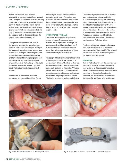
Fig. 3<br />
Fig. 4<br />
Fig. 5: VITA Akzent LC was chosen as the composite stains<br />
Fig. 6: A view of the completed crowns that took 90mins to produce<br />
40 DENTAL ASIA JANUARY / FEBRUARY <strong>2024</strong>


